|
|
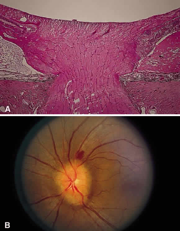
| Fig. 30. A. Photomicrograph of an optic disc with papilledema. There is edema of the disc surface, some engorgement of the vessels, and lateral displacement of the photoreceptor elements, which results in enlargement of the blind spot in papilledema. B. Early papilledema in a patient with pseudotumor cerebri. The disc margin is blurred, the surface slightly elevated, and a small hemorrhage is present superiorly. (A, courtesy of Ralph C. Eagle Jr, MD, Philadelphia, PA) |