|
|
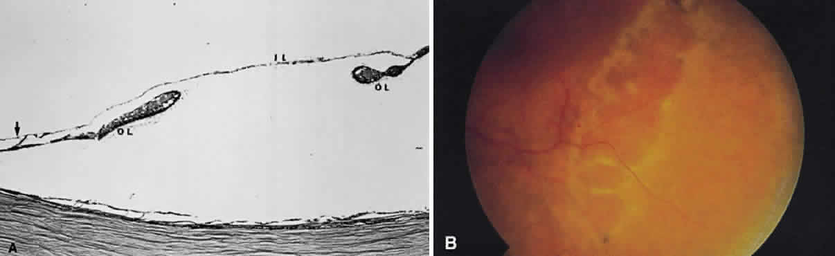
| Fig. 60. A. Photomicrograph of a retinoschisis cavity with a hole in the outer layer (OL). The inner layer (IL) is intact. Typical peripheral cystoid degeneration is present (arrow). B. Outer wall holes in a patient with senile retinoschisis. Notice the pocked marked appearance of the outer wall layer peripheral to the holes. (B, courtesy of William Benson, Philadelphia, PA, Wills Eye Atlas of Ophthalmology. Lippincott-Raven Fig 4-58, 1996) |