|
|
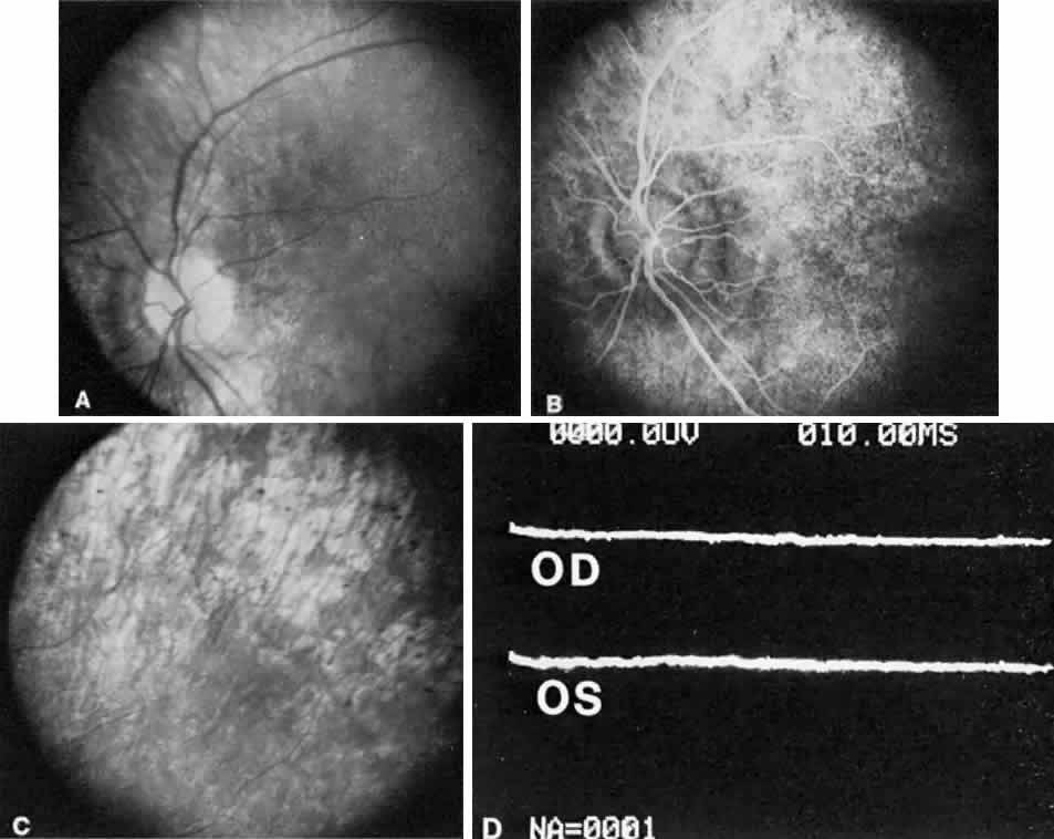
| Fig. 25. A. Fundus of a 4-year-old child with Leber's congenital amaurosis. Diffuse pigmentary changes are present in the macula, the optic disc is slightly pale, and the retinal arteries are somewhat narrowed. The child has nystagmus, and there was no central fixation in either eye. B. Fluorescein angiogram of A reveals a pattern of mottled hyperfluorescence better delineating the posterior-pole retinal pigment epithelium disturbance. C. Peripheral fundus photograph shows more marked pigmentary changes. D. The electroretinogram is flat in each eye. (Courtesy of Dr. William Tasman) |