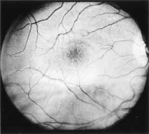
Fig. 2
X-linked juvenile retinoschisis. Red-free photography is well suited for demonstrating both the central foveal schisis and the fine radiate plication of the internal limiting membrane.