|
|
|
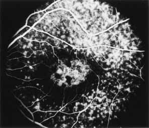
Fig. 15 Stargardt's
disease. Fluorescein angiography in arteriovenous phase shows an oval zone
of retinal pigment epithelium (RPE) transmission window defect within the
central macula. The hypofluorescent spots correspond to pigmentations developing
within this area (see Fig. 14 |