|
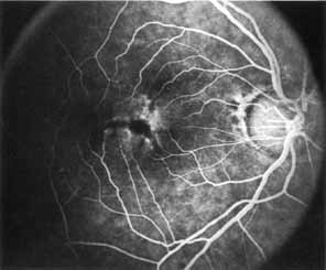
Fig. 30 Dominant slowly
progressive macular dystrophy. Fluorescein angiography in midarteriovenous
phase of patient seen in Fig.
28 demonstrates a fluorescent pattern intermittent between
foveomacular vitelliform dystrophy: adult type (see Fig.
27) and macroreticular dystrophy (see Fig.
34). |