|
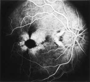
Fig. 33 Butterfly-shaped
pigment dystrophy of the fovea. Fluorescein angiography in mid arteriovenous
phase of patient seen in Fig.
32 shows clearly reticular or propeller-like hypofluorescent
pattern corresponding to the yellow and yellow-gray deposition material.
Pigment epithelial transmission hyperfluorescence is present within this
pattern. |