|
|
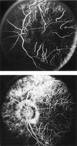
| Fig. 40 Central areolar choroidal dystrophy. Top. Early venous phase. Bottom. Midvenous phase. Fluorescein angiography in arteriovenous phase reveals the diffuse epithelial and choriocapillary atrophy and lack of choriocapillary filling and hyperfluorescence within the area of macular involvement. Elsewhere, the choriocapillaris does show filling, although it is not entirely normal. |