

|
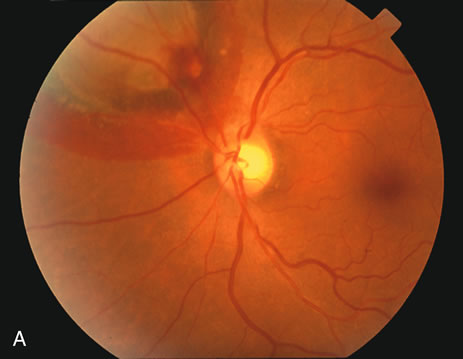
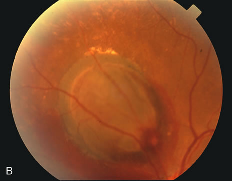
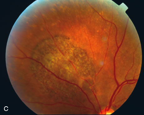
| Fig. 4. A. Color fundus photograph of a left eye with a 300-micron macroaneurysm along the superonasal arcade with a localized area of thick subretinal hemorrhage. B. The macroaneurysm and subretinal hemorrhage were not treated. C. After several months, the subretinal hemorrhage resorbed with visual acuity of 20/20. |