|
|
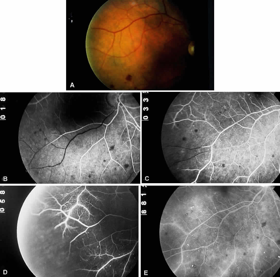
| Fig. 4. A. Fundus photograph of an eye with ocular ischemic syndrome showing features of venous dilation, attenuated arterioles, and midperipheral intraretinal hemorrhages. B. Fluorescein angiogram at 18 seconds, displaying a visible leading edge of fluorescein dye within the inferotemporal arteriole. C. Numerous microaneurysms along the inferotemporal arcade. At 33 seconds, the arteriolar dye front is still apparent. D. Retinal capillary nonperfusion and microaneurysms at 58 seconds. E. Prominent arterial staining in the late phase of the angiogram. |