|
|
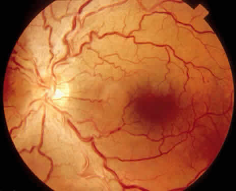
| Fig. 1. Fundus photograph demonstrating vasoconstrictive and early sclerotic changes seen in hypertensive retinopathy, including areas of diffuse arteriolar narrowing, sinusoidal tortuosity, and copper wire appearance. There also are arteriovenous crossing changes, with tapering of the veins and increased arteriolar branching angles. |