|
|
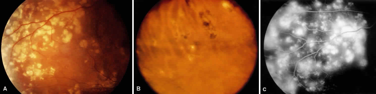
| Fig. 9. Fundus photograph showing retinal changes secondary to hypertensive choroidopathy. A. Elschnig's spots are ischemic infarcts of the RPE that appear in the posterior pole. B. Siegrist's spots are ischemic infarcts of the RPE that appear at the equator. They become hyperpigmented as the RPE repairs the injury. C. Fluorescein angiogram reveals early hyperfluorescence caused by disruption of the RPE blood-retinal barrier in areas of the Elschnig's spots. |