|
|
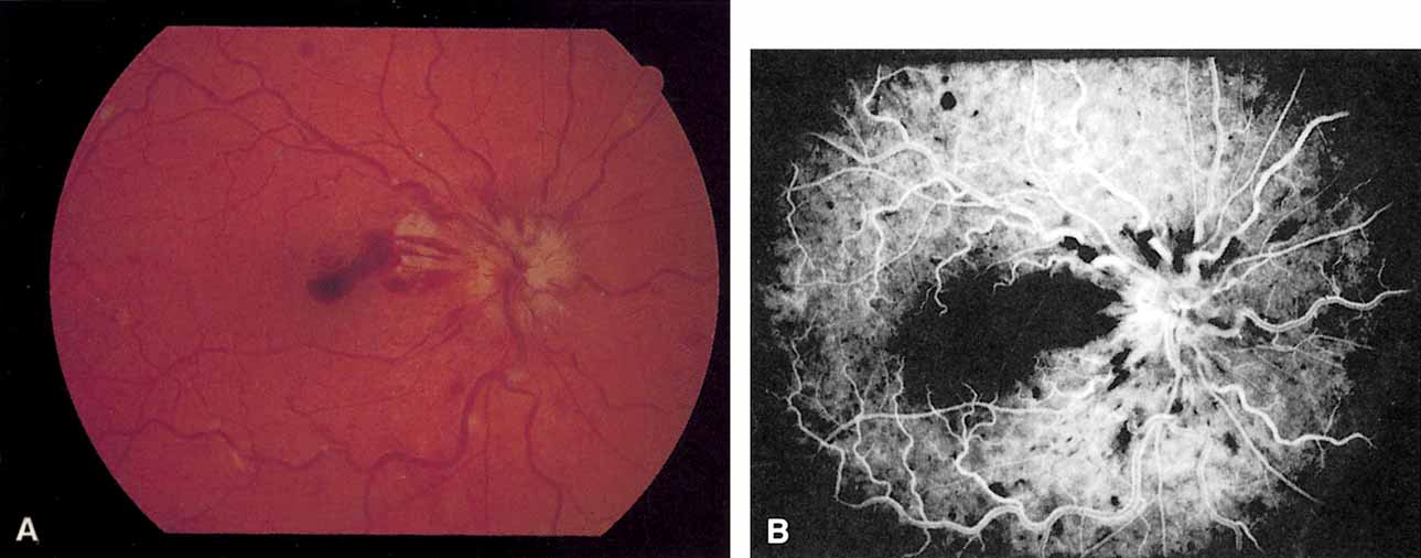
| Fig. 2 A. Nonischemic central retinal vein occlusion. Note venous engorgement, dot, blot, and flame-shaped hemorrhages, blurring of disc margins, and a hemorrhage overlying the macula. B. Fluorescein angiogram reveals mild venous engorgement and tortuosity with virtually no capillary nonperfusion. |