|
|
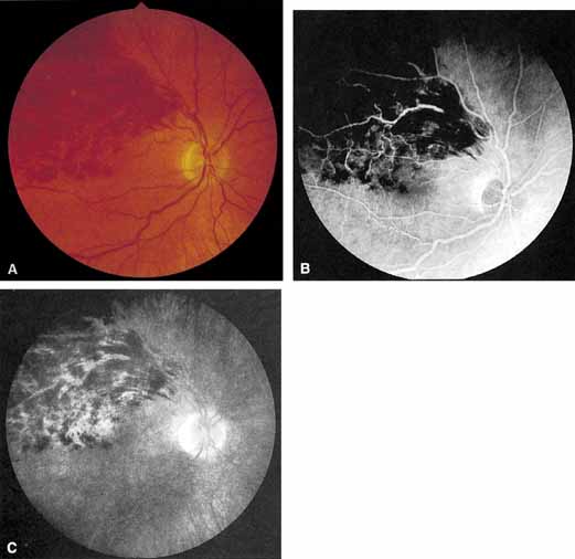
| Fig. 8 A. Nonischemic superior temporal branch retinal vein occlusion. The visual acuity is reduced because of mild macular edema. B and C. The intravenous fluorescein angiogram shows the nonischemic nature of this occlusion and macular edema in the late stage of the angiogram. |