|
|
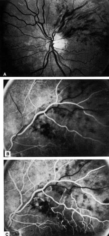
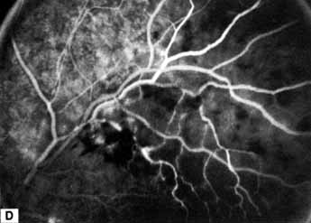
| Fig. 9 A. Acute branch retinal vein occlusion with apex located at an arteriovenous crossing point at the superior disc margin. Striate hemorrhages are predominant. B. Early venous phase fluorescein angiogram in which the superior temporal artery is filled with fluorescein and early venous return is present in an unobstructed branch of the superior retinal vein. No fluorescein has entered the occluded vein segment. C. Midvenous phase. Fluorescein has still not entered the occluded branch. D. Late venous phase. Fluorescein now fills the occluded vein segment and can be seen draining into the main superior trunk beyond arteriovenous crossing. |