
|
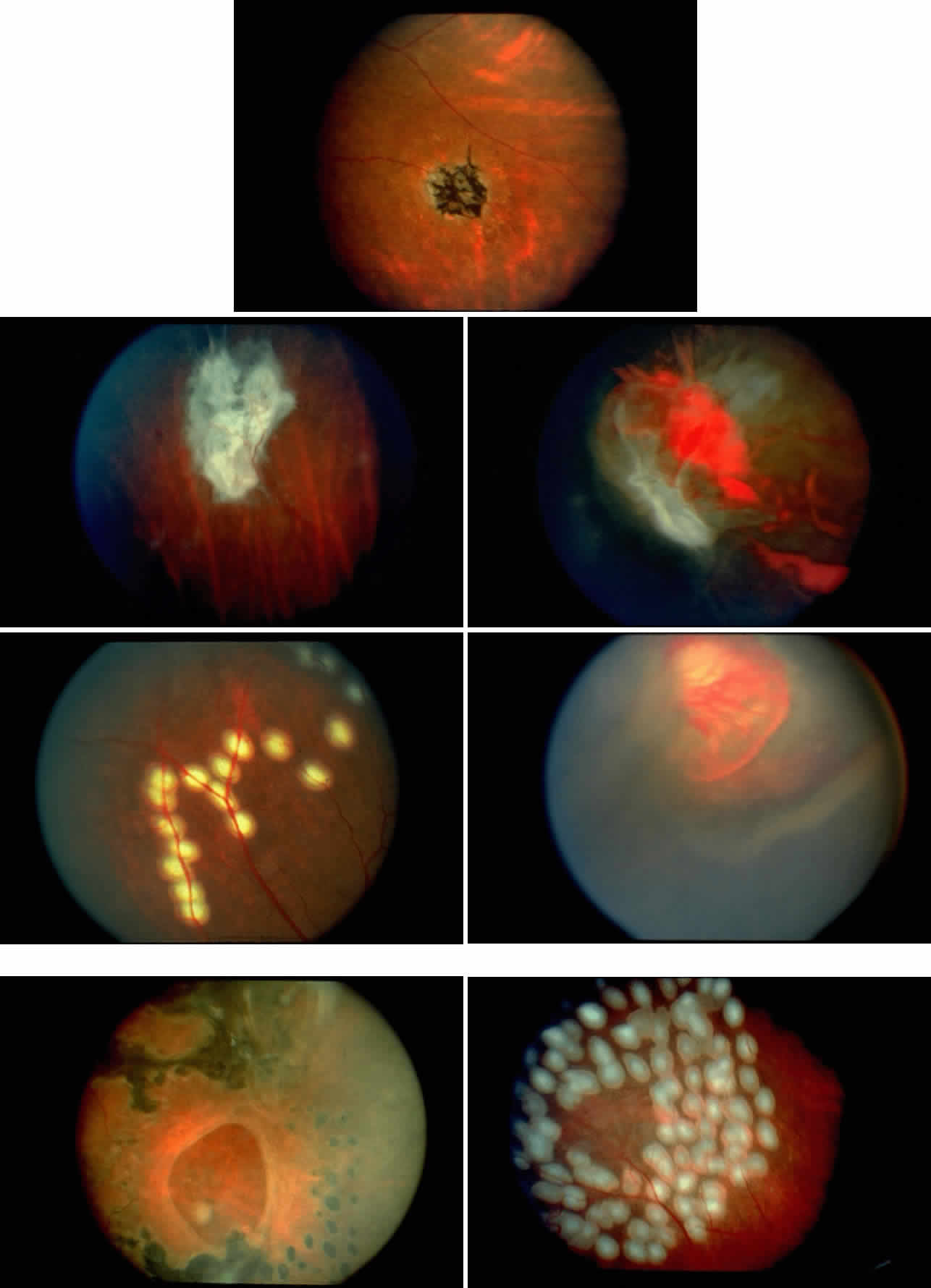
| Color Plate 2. A. Black sunburst with periarteriole pigment cuff. B. Elevated sea fan neovascularization with white fibroglial mantle. C. Untreated sea fan with localized and diffuse vitreous hemorrhage. D. Feeder vessel photocoagulation immediately after treatment, demonstrating segmented vessels over photocoagulation spots. E. Chorioretinal and choriovitreal neovascularization after feeder vessel photocoagulation. F. Peripheral retinal hole in a patient who was treated with feeder vessel photocoagulation applied to two adjacent sea fans. Scatter photocoagulation was placed around the hole. G. Scatter photocoagulation surrounding a perfused sea fan shown immediately after treatment. (D and G; Gagliano DA, Rabb MF: Sickle cell retinopathy. In Cowan CL (ed): Mediguide to Special Problems in Ophthalmology. New York, Lawrence Della Corte Publications, 1991.) |