|
|
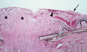
| Fig. 13. Histopathology of papilledema. Note the marked swelling and elevation of the nerve fiber bundles in the optic disc (asterisks), lateral displacement of the peripapillary retina from the disc margin (bracket), nerve fiber layer hemorrhage (arrowhead), and subhyaloid hemorrhage (arrow). |