|
|
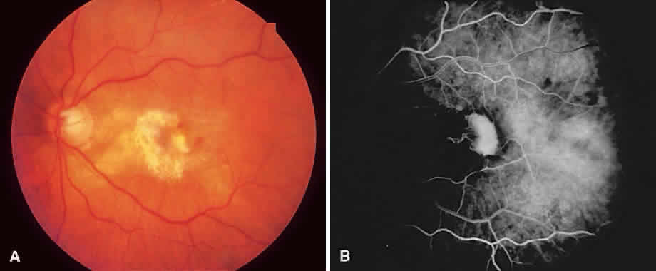
| Fig. 11. A. Atrophic laser scar with hemorrhage and subretinal fluid at the temporal border signals recurrent subretinal neovascularization. B. Early-phase angiogram shows a racquet pattern feeder vessel giving rise to the recurrent choroidal neovascularization complex. |