|
|
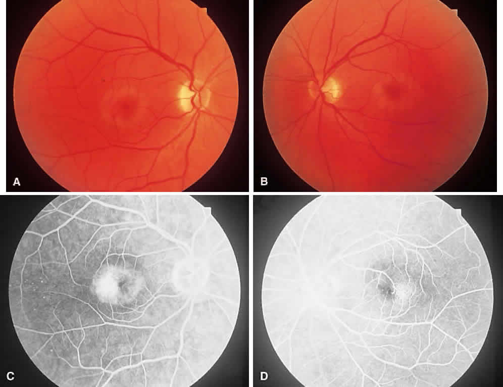
| Fig. 27. Group 2A idiopathic juxtafoveolar retinal telangiectasia. A & B. Both eyes show involvement. A retinal pigment plaque is noted in the right macula, denoting more extensive involvement. Note the surrounding retinal atrophy. C & D. Angiogram shows leakage greater in the right eye than the left. Note the transmitted fluorescence from foveal atrophy. |