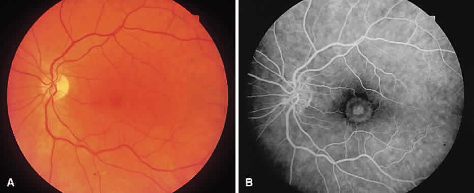
Fig. 30.
A.
Stage 3 macular hole with cuff of subretinal fluid.
B.
Angiography reveals transmitted fluorescence at the hole and leakage into the subretinal space corresponding the cuff of fluid.