|
|
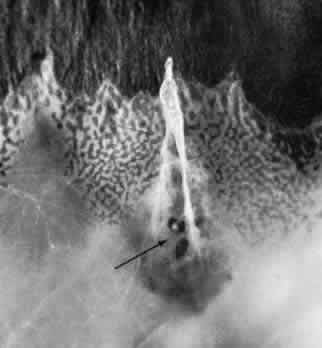
| Fig. 40. Zonular-traction tuft of the peripheral retina. Tuft is drawn at an acute angle from the retinal surface toward the ciliary body and shows microcystic degeneration anteriorly. Posteriorly, the tuft splays; the retina at its base shows marked trophic change, including three full-thickness holes (arrow). (× 20.) |