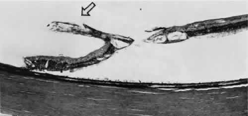
Fig. 41.
Zonular-traction tuft (
arrow
) drawn at an acute angle from the retinal surface toward ciliary body with dense-staining glial cells along surface. Base of tuft is microcystic and has full-thickness trophic hole. (Hematoxylin-eosin; × 150.)