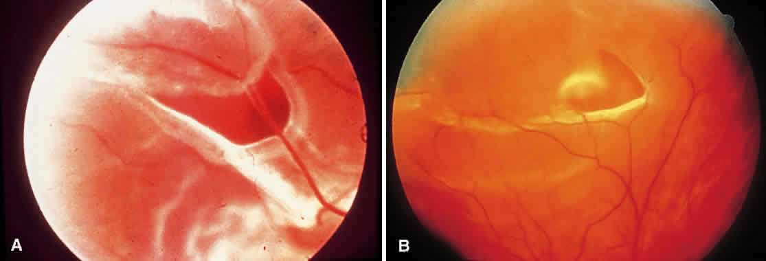
Fig. 2.
A.
Flap (horseshoe tear) with a rolled posterior edge. Bridging vessels can be seen.
B.
A flap tear associated with a small rhegmatogenous retinal detachment. (
A
courtesy of William E. Benson, MD, Philadelphia, PA.)