|
|
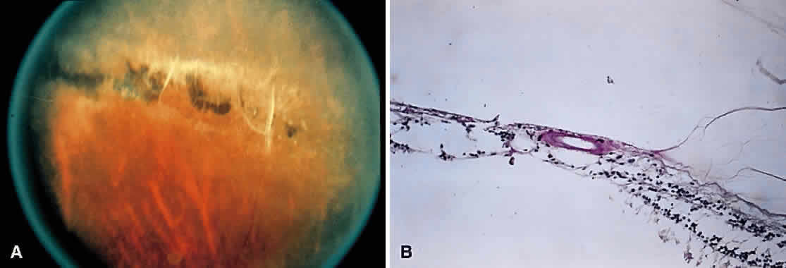
| Fig. 5. A. Lattice degeneration with pigmentation. Hyalinized blood vessels are seen in the lattice. B. Photomicrograph of lattice degeneration demonstrating focal thinning of the inner retinal layers and associated liquefaction of the overlying vitreous with vitreous attachments to the margins. (Courtesy of William Tasman, MD, Philadelphia, PA.) |