|
|
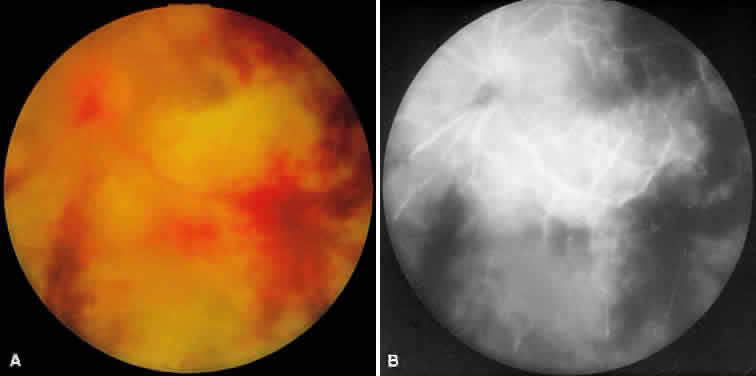
| Fig. 7. A. Active ARN with diffuse retinal necrosis progressing into the macula and areas of retinal hemorrhage. The hazy view is secondary to vitreous inflammation. B. Fluorescein angiogram in the venous phase of active ARN syndrome in this eye reveals peripheral vascular nonperfusion, as well as optic nerve and perivascular hyperfluorescence. |