|
|
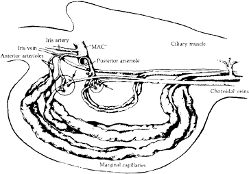
| Fig. 7. Diagrammatic representation of Fig. 6 demonstrating the system of anterior and posterior arterioles that enter each ciliary process, ultimately draining into a set of choroidal veins. (From Morrison JC, Van Buskirk EM, Freddo T. Anatomy, microcirculation and ultrastructure of the ciliary body. In: Ritch R, Shields MB, Krupin T (eds). The Glaucomas. St Louis: CV Mosby, 1989.) |