

|
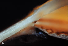
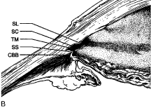
| Fig. 18. Macroscopic photograph (a) and corresponding sketch (b) identifying structures visible in a meridional section of the normal anterior chamber angle. The anterior chamber is artificially deepened because of posterior sagging of the iris following removal of the crystalline lens. The heavily pigmented region corresponds to the posterior or “filtering” meshwork. SL, Schwalbe's line; SC, Schlemm's canal; TM, trabecular meshwork; SS, scleral spur; CBB, ciliary body band. (From Freddo T: Ocular anatomy and physiology related to aqueous production and outflow. In: Fingeret M, Lewis T (eds). Primary Care of the Glaucomas. 2nd ed. New York: McGraw-Hill, 2001.) |