|
|
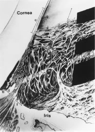
| Fig. 21. Drawing of the aqueous outflow apparatus and adjacent tissues shown from two perspectives—a gonioscopic view and the view obtained from meridional sections. The positions of the dark bands correspond to the positions of the pigmented bands seen in the goniophotograph in Fig. 20. The uppermost dark band covers Schwalbe's line (g) and the transition of the corneal endothelium and the trabecular endothelium (j). The uppermost light band corresponds to the anterior or “nonfiltering” meshwork. Note that this portion of the meshwork does not lead to Schlemm's canal (a). The next dark band corresponds to the posterior or “filtering” meshwork that does lead to Schlemm's canal. The next light band corresponds to the scleral spur (d), and the lowest dark band corresponds to the “ciliary body band.” CB, Ciliary body; i, longitudinal bundle of the ciliary muscle; h, iris process. (From Hogan M, Alvarado J, Weddell J: Ciliary body and posterior chamber. In: Histology of the Human Eye. Philadelphia: WB Saunders, 1971, p 137.) |