|
|
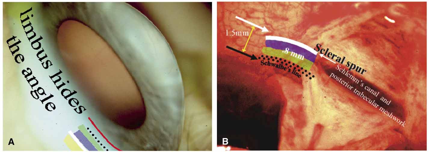
| Fig. 9 Peripheral anterior chamber depth: anatomic correlates and why estimates of limbal chamber depth (van Herick) are not a substitute for gonioscopy. A. The loss of transparency at the corneoscleral limbus prevents a direct view of the underlying chamber angle. The projected surface locations of the outflow structures are noted to demonstrate how the limbus normally conceals the angle structures. A thorough understanding of this area is crucial especially during angle surgery and in classifying the glaucomas. The projected structures in order from anterior to posterior include: red, corneal limbus, conjunctival epithelium ends at periphery of Bowman's layer of the cornea; dotted black line, approximate end of Descemet's membrane and beginning of outflow system typically seen during gonioscopy as Schwalbes' line; green zone, anterior trabecular outflow; blue zone, posterior trabecular outflow; white, approximate location of scleral spur, posterior limit of limbus and trabecular outflow system; and yellow zone, ciliary body band and beginning of uveoscleral outflow. B. Because of the transition of orderly collagen fibers into coarse scleral fibers at the corneoscleral junction, there is a loss of transparency at the limbus and the location of angle structures is unknown. Guess the location of the scleral spur in this intraoperative photograph. You would probably guess the black arrow, but it is actually the white arrow. This is proved by the layer-by-layer dissection that is required during a nonpenetrating procedure. During a deep sclerectomy procedure where the deep scleral flap is retracted, the underlying angle structures become visible. The scleral spur, white arrow, is seen well posterior to the corneal limbus, black arrow. Most ophthalmologists do not realize how far posterior the canal and meshwork may be located. The opposite may hold true with the spur situated very close to the corneal limbus. During this deep sclerectomy, the dissection of the deep flap reveals the scleral spur, floor of Schlemm's canal, and underlying posterior trabecular meshwork. It is rare for ophthalmologist to see this layer-by-layer dissection of the limbus and note how it correlates with what they see at the slit lamp. The point of the discussion is that the location of the scleral spur is highly variable and the van Herick test is unable to compensate for this, thus the reason gonioscopy is essential. (Figure continues.) |