
|
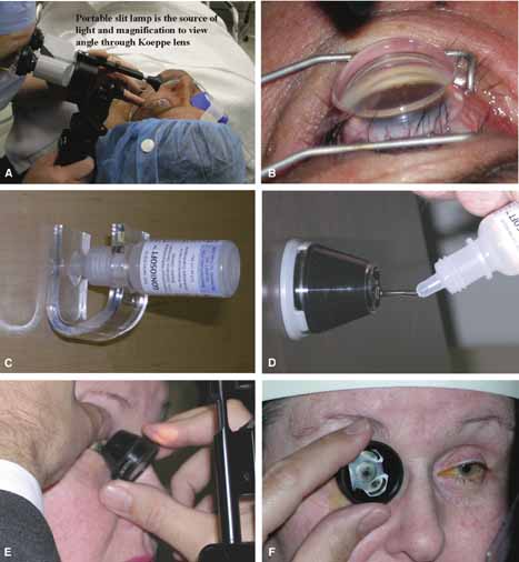
| Fig. 12 Gonioscopy techniques. A. Koeppe gonioscopy requires a hand-held slit lamp to view the angle, a lid speculum to assist in inserting the device, an appropriately sized Koeppe lens, and a saline bridge. The slit lamp is held in the examiner's dominant hand. Simultaneously the nondominant hand acts as a proprioceptive bridge to steady the slit lamp and keep the correct distance to the eye. B. Angle view through medium-sized Koeppe lens. The oniophotograph reveals intermittent PAS secondary to chronic trabeculitis. C. The Goldmann lens comes in various sizes in order to fit into the palpebral fissure area. Store the methycellulose viscous material inverted to avoid the instillation of air bubbles onto the contact lens. D. Apply the goniogel to the lens in a bubble-free fashion. E. Tell the patient that a contact lens will be placed on their eye in order to view the drainage area. The patient should be seated the at the slit lamp so that the eye is level with the positioning marks on the headrest. The upper lid is grasped and the patient instructed to look up slightly. The contact lens portion of the prism is tilted up to ease the insertion of the lower pole of the Goldmann lens into the lower fornix and onto the globe. F. As the lens is rotated into position, the patient is asked to look forward to bring the cornea into contact with the Goldmann and the upper lid is simultaneously released. There are variations of this technique; the examiner must find what works best for him. (Figure continues.) |