|
|
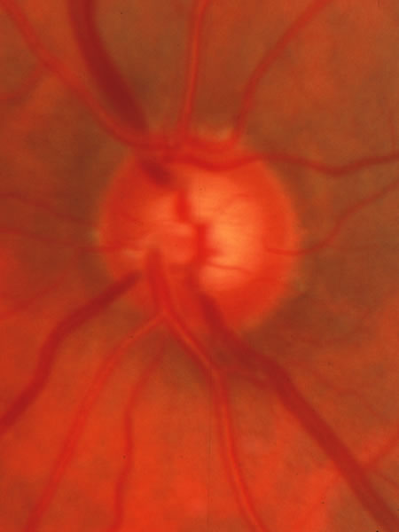
| Fig. 19. Same disc as Figure 18 after 5 years of elevated intraocullar pressure. The horizontally oriented venous tributary that crosses the disc margin in the 1:00 position has less tissue between it and the cup than in Figure 18, showing the beginning of glaucomatous tissue loss. Even without the baseline photograph, a suspicion of early glaucoma may be recognized by the fact that the neuroretinal tissue in the upper sector is not thicker than the rim in the nasal sector the way it should be. |