|
|
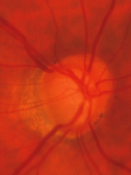
| Fig. 40. Same eye photographed in 1998, after progression of the cupping and visual field loss. Continued thinning of the neuroretinal rim is evident in nearly every part of the circumference and the course of the veins across the inferior border of the disc show the deeper excavation. This progression would not likely be evident if only disc drawings were used to monitor the disc status. Stereoscopic fundus photographs of other forms of imaging are discussed in Chapter 48A are needed. |