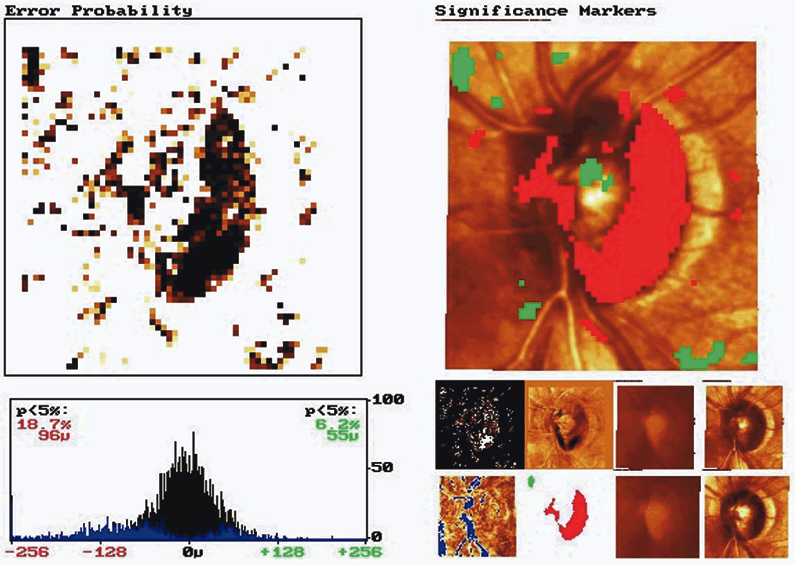
Fig. 10.
Probablity map analysis: glaucomatous optic disc. This probability map compares the baseline and follow-up examination of a glaucoma patient over 5 years. The red markers indicate diffuse glaucomatous progression of the neuroretinal rim.