
|
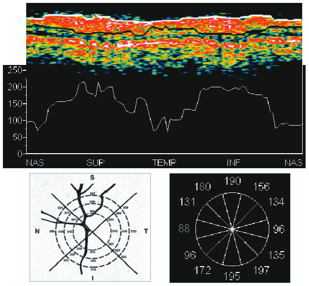
| Fig. 17. Optical coherence tomography: healthy eye. In this example of a healthy eye, the anterior and posterior highly reflecting layers can be seen, representing the retinal nerve fiber layer (RNFL) and retinal pigment epithelium (RPE), respectively. The inferotemporal and superotemporal nerve fiber bundles are evident as localized thickenings in both the RNFL and the retina. |