|
|
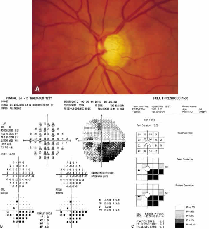
| Fig. 23. Full threshold perimetry and frequency doubling technology. A. Disc photo of the left eye demonstrating a superior notch with inferior thinning. Both Humphrey visual field (B) and frequency doubling technology field (C) show predominantly inferior defects. |