|
|
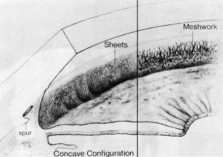
| Fig. 2 Concave iris insertion in trabeculodysgenesis. Superficial iris tissue wraps around the angle recess and covers the internal surface of the trabeculum. This may take the form of dense sheets (left) or an arborizing network (right). This is different from the small processes seen in the anterior iris insertion. (Hoskins HD Jr, Shaffer RN, Hetherington J: Anatomical classification of the development of glaucomas. Arch Ophthalmol 102:1331, 1984. Copyright © 1984, American Medical Association.) |