
|
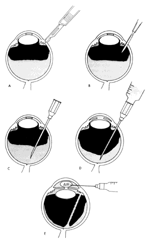
| Fig. 27. Chandler's vitreous operation. A. After a conjunctival incision, a radial scleral incision is centered 3.5 mm behind the external limbus. Diathermy (not shown) is then applied around the scleral wound. B. A Wheeler knife is used to pierce the uvea and enter the vitreous cavity. The knife is kept away from the lens by aiming it toward the optic nerve head. C. An 18-gauge needle is inserted 12 mm into the eye. (Hemostat guard to control needle depth is not shown.) D. A syringe is attached and 1 to 1.5 mL of fluid is aspirated. E. A very large air bubble is placed in the anterior chamber to deepen it to abnormal depth. The small air bubble shown will be enlarged by continued injection. (Simmons RJ: Malignant glaucoma. Br J Ophthalmol 56:263, 1972) |