|
|
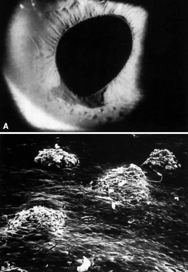
| Fig. 3. Iris nodules in ICE syndrome. A. Multiple nodules stud flattened area of iris stroma inferiorly, adjoining distorted pupil and iris pigment epithelial ectropion. Stromal effacement and iris nodules are a clinical marker for iris endothelialization. B. Scanning electron microscopy (SEM) of anterior iris surface in area of clinically effaced stroma shows multiple iris nodules surrounded by confluent sheet of corneal endothelial cells. Characteristic iris nodules appear to be formed by the encirclement of knuckles of iris stroma by the proliferating corneal endothelium. (SEM, x200) (Courtesy of Ralph C. Eagle Jr, MD) |