Chapter 22
Vascular Anomalies of the Fundus
CAROLINE R. BAUMAL, JAY S. DUKER and GARY C. BROWN
Main Menu   Table Of Contents

Search

CHORIOVAGINAL VEIN
PERSISTENT HYALOID ARTERY
PREPAPILLARY LOOPS
HEREDITARY RETINAL ARTERY TORTUOSITY
INHERITED RETINAL VENOUS BEADING
RETINAL MACROVESSELS
ARTERIOVENOUS COMMUNICATIONS
IDIOPATHIC JUXTAFOVEOLAR RETINAL TELANGIECTASIS
CONGENITAL RETINAL TELANGIECTASIS (COATS' DISEASE)
CAPILLARY HEMANGIOMA OF THE RETINA
VASOPROLIFERATIVE TUMORS OF THE FUNDUS
CAVERNOUS HEMANGIOMA OF THE RETINA
CHOROIDAL HEMANGIOMA
IDIOPATHIC POLYPOIDAL CHOROIDAL VASCULOPATHY
IDIOPATHIC RETINAL VASCULITIS, ANEURYSMS AND NEURORETINITIS
VARIX OF THE VORTEX VEIN
REFERENCES

Choroidal and retinal vascular anomalies may be associated with systemic disorders. Accurate diagnosis is therefore critical. In most instances, careful ophthalmoscopic examination is sufficient, In some cases, ancillary tests such as intravenous fluorescein angiography (IVFA), indocyanine green (ICG) angiography, ultrasonography, computed tomography (CT) and magnetic resonance imaging (MRI) are necessary either to confirm the diagnosis or to rule out associated systemic disorders.
Back to Top
CHORIOVAGINAL VEIN

A choriovaginal vein is an uncommonly recognized, unusually large choroidal vessel located within the posterior pole, The choriovaginal vein passes from the choroid through the sclera at the margin of the optic disc to terminate in the venous plexus of the pial sheath of the optic nerve.1 This establishes an indirect communication between the choroidal and retinal venous systems through the pial branches of the central retinal vein.2 It is generally seen temporal to the optic nerve and is predominantly noted in lightly pigmented, myopic fundi1,3 (Fig. 1). This entity was described in a case of trisomy 13 with congenital glaucoma, although it is not typically associated with any systemic abnormalities.4

Fig. 1. A choriovaginal vein (posterior vortex vein) situated just temporal to the optic disc in a highly myopic eye. These unusually located choroidal vessels are believed to be tributaries of the ophthalmic vein. A Fuch's spot is present in the fovea.

Back to Top
PERSISTENT HYALOID ARTERY
The hyaloid vessels develop around the fifth week after conception. The hyaloid artery provides the bulk of blood supply to the developing posterior segment and lens during late embryologic and early fetal development. Its branches include the tunica vasculosa lentis and the vasa hyaloidea propia. Spontaneous regression of the fetal hyaloid artery commences at the start of the third trimester (28 weeks),5–7 Ultrasound examination during pregnancy typically demonstrates the hyaloid artery in fetuses of 20 weeks' gestational age or younger, whereas its presence on ultrasound in the mid-third trimester is uncommon in healthy fetuses.8

A persistent hyaloid artery represents incomplete regression and is noted clinically as a single threadlike vessel emanating from the optic disc. It travels in a sinuous course anteriorly through the vitreous cavity within Cloquet's canal. Persistent hyaloid arteries can traverse the entire length of the vitreous cavity to insert on the posterior capsule of the lens (Fig. 2). When all but the anterior lens insertion of the hyaloid artery regresses, the remaining focal posterior lens capsule opacity is called Mittendorfs dot. At birth, up to 95% of premature infants have hyaloid artery remnants. Such remnants also have been noted in 3% of full-term infants.6 Persistent hyaloid arteries may be filled with blood. When they are blood filled, vitreous hemorrhage may occur. This can be spontaneous, precipitated by trauma or by a posterior vitreous detachment.9,10 It has been postulated that rapid eye movements during sleep produce traction, leading to rupture of a freely floating hyaloid artery in young individuals.11 Other ocular conditions that may be associated with a persistent hyaloid artery include strabismus, cataract, amblyopia, and nystagmus.10 A persistent hyaloid artery typically does not need treatment, but recurrent vitreous hemorrhage secondary to this condition may require vitrectomy. Also, a persistent, patent hyaloid artery may complicate attempts at surgical repair in infants with the condition called persistent hyperplastic primary vitreous or with retinal detachment caused by retinopathy of prematurity.12

Fig. 2. Slit-lamp view of a persistent hyaloid artery inserting on the posterior lens capsule.

Back to Top
PREPAPILLARY LOOPS
Prepapillary vascular loops were first recognized by Leibrich in 1871.13 These congenital vascular anomalies extend into the vitreous cavity. They appear as corkscrew, spiral, or hairpin vessels overlying the optic disc and are contiguous with the normal retinal circulation (Fig. 3A). They can arise from the central retinal vessels, a branch retinal vessel, or a cilioretinal vessel,6,7,14 About one third of prepapillary loops are encased in a fibroglial sheath, and they are present bilaterally in approximately 10% of affected persons,6 Eyes containing a prepapillary loop have an unusually high prevalence of associated cilioretinal arteries. On ophthalmoscopic examination, prepapillary loops may pulsate. IVFA has shown that 95% of prepapillary loops are arterial in origin.6,15

Prepapillary loops were once believed to be remnants of the hyaloid artery, It is now thought that they represent developmental anomalies that occur when mesenchymal cells destined to differentiate into mature retinal vessels proliferate within Berg-meister's papilla.16 Histopathologic study has demonstrated communication of a prepapillary loop with the retinal arterial system.16 The prepapillary loop was supported by a connective tissue sheath that was lined by a cellular lamina continuous with the inner retina.

Fig. 3. A. A prepapillary loop derived from the central retinal artery. Notice the fibrous sheath. B. Branch retinal artery occlusion associated with a prepapillary loop.

The major complication occurring with prepapillary loops is retinal arterial obstruction within the territory supplied by the loop (see Fig. 3B). This is believed to occur secondary to turbulent flow, endothelial damage, and subsequent thrombus formation.15 Vitreous hemorrhage, amaurosis fugax, hyphema, and retinal venous macrovessel also have been reported.15,17–20 There are no associated systemic abnormalities. Two studies report a familial occurrence of prepapillary loops, although a definite hereditary pattern has not been identified.21,22

Back to Top
HEREDITARY RETINAL ARTERY TORTUOSITY
Hereditary retinal artery tortuosity is a rare, congenital, autosomal dominantly inherited condition characterized by extreme tortuosity of the retinal arteries. Patients with this entity may present acutely with visual loss from a superficial macular hemorrhage that occurs spontaneously or after relatively minor trauma.14,23–25 Vision typically returns to normal as the retinal hemorrhage clears. The artery tortuosity is most marked in the macular region, whereas the retinal veins remain normal (Fig. 4). The vascular tortuosity progressively increases with age.25 On IVFA, the tortuous retinal arteries are competent, and there is no microvascular cause for the macular hemorrhage. Histologic study by light microscopic examination in one case failed to reveal any structural abnormality in the arteriolar wall.23 No systemic vascular abnormality has been detected in these patients.26 The other syndrome that typically produces isolated retinal arteriolar tortuosity without venous involvement is coarctation of the aorta.27 It may be reasonable to examine family members for arteriolar tortuosity before subjecting an affected individual to an extensive medical evaluation. Hereditary retinal artery tortuosity should be differentiated from other systemic diseases that can induce retinal vascular tortuosity (affecting both the arteries and veins) including hypoxic retinal syndromes (e.g., sickle cell disease, congenital heart disease), leukemia, dysproteinemias, familial dysautonomia, mucopolysaccharidosis, and Fabry's disease.14

Fig. 4. Hereditary retinal artery tortuosity in a 15-year-old asymptomatic boy, Notice the normal-appearing retinal veins. The patient's father had a similar retinal vascular pattern.

Back to Top
INHERITED RETINAL VENOUS BEADING
Meredith reported a family in whom some of the members presented with prominent bilateral beading and segmentation of their retinal veins.28 Associated findings included beading of the conjunctival veins, retinal capillary nonperfusion, microaneursym formation, altered vascular permeability with lipid exudation, retinal edema, retinal neovasvularization, and vitreous hemorrhage. The family's pedigree was consistent with autosomal dominant inheritance. These retinal vascular changes may represent an unusual manifestation of renal disease, since two of the five patients concurrently had Al-port's syndrome, A second reported pedigree showed no evidence of renal disease, although affected members did display a relative neutropenia on peripheral blood examination.29
Back to Top
RETINAL MACROVESSELS
A congenital retinal macrovessel is an aberrant, enlarged branch retinal vessel that supplies or drains the macular area, crossing the horizontal raphe (Fig. 5A). They usually are veins. The condition typically is unilateral, benign, and ophthalmoscopically stable. This entity often is detected on routine examination.14 Visual acuity is typically unaffected, although concomitant foveal cysts have been noted.30 IVFA may reveal delayed drainage of dye from the macrovessel, associated microvascular abnormalities, and retinal capillary nonperfusion30 (see Fig. 5B). On occasion, retinal macrovessels may be accompanied by similar vascular anomalies affecting the conjunctiva and mouth.14

Fig. 5. A. Superotemporal retinal venous macrovessel traversing the horizontal raphe and a foveal cyst. B. IVFA demonstrates the retinal venous macrovessel and foveal cyst. There is no extravascular leakage.

The retinal arteries and the accompanying capillary bed are relatively normal in eyes with congenital retinal macrovessels. This is in contrast to congenital retinal arteriovenous communications, which do not feature a normal intervening capillary bed.

Back to Top
ARTERIOVENOUS COMMUNICATIONS
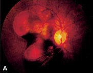
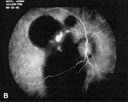
Congenital retinal arteriovenous communications are abnormally dilated retinal vessels that are accompanied by an abnormal or absent capillary system, resulting in a more direct connection between afferent and efferent vessels. They are unilateral and involve either single or multiple sites with a predilection for the papillomacular area and the superotemporal quadrant.31 Both sexes are equally affected. Archer and colleagues divided these communications into three types based on size of the affected vessels and condition of the intervening capillary bed.32 Type I is a typical retinal macrovessel associated with an intervening capillary bed containing abnormally dilated vessels. Archer's type II retinal arteriovenous communication consists of a dilated and tortuous artery and vein directly connected, without an intervening capillary bed. Type III arteriovenous communication is an exaggerated version of type II, with the artery and vein so aberrant and intertwined that no distinction between afferent and efferent vessels can be made on ophthalmoscopic examination (Fig. 6A). Type II and type III malformations also are referred to as racemose, cirsoid or arteriovenous aneurysms, although they are not true aneurysms.14,33

Fig. 6. A. Archer's grade III retinal arteriovenous communication (racemose aneurysm) in a patient without the Wyburn-Mason syndrome. Visual acuity was 20/200 (6/60). B. Superotemporal branch retinal vein occlusion associated with a grade III retinal arteriovenous communication (racemose aneurysm), (A and B, courtesy of Jerry A. Shields, MD and Carol L. Shields, MD)

When arteriovenous communications are large, visual acuity can be severely affected, and the optic nerve tissue can be completely replaced by the anomalous vessels. Vascular sheathing and retinal pigmentary degeneration may occur. Patients with more severe retinal involvement are more likely to have cerebral or periorbital arteriovenous abnormalities, which comprises the Wyburn-Mason syndrome.31

The clinical appearance of these lesions may change over time.34,35 Complications associated with this vascular abnormality include intraretinal hemorrhage, vitreous hemorrhage, aneurysm formation, neovascular glaucoma, macular hole, retinal artery occlusion, and branch or central retinal venous occlusion (see Fig. 6B).36–40 It is hypothesized that retinal arteriovenous communications are associated with localized decreased retinal arterial pressure, increased retinal venous pressure, increased turbulence of blood flow, and decreased perfusion of adjacent retinal tissues to account for these complications.36 IVFA may show rapid dye transit through the arteriovenous communication. There may be adjacent microvascular abnormalities or areas of capillary nonperfusion. Extravascular leakage from these lesions is uncommon. Congenital retinal arteriovenous communications should be distinguished from those acquired secondary to carotid occlusive disease associated with retinal ischemia.41 No successful treatment of these lesions has been reported. Laser photocoagulation may be warranted in some cases with exudative macular involvement or neo-vascular glaucoma.14,37

A racemose or cirsoid retinal arteriovenous communication may represent the ocular manifestation of Wyburn-Mason syndrome, which is one of the phakomatoses.33 This syndrome consists of unilateral retinal arteriovenous communication in conjunction with an ipsilateral arteriovenous malformation of the midbrain.42 The presence of a concomitant systemic arteriovenous communication correlates directly with the grade of retinal arteriovenous communication. Most grade I and II arteriovenous communications appear to represent isolated retinal vascular anomalies.31 Unlike the other phakomatoses, the hallmark ocular lesion of Wyburn-Mason syndrome is not a true hamartoma but is better classified as a hamartia.33 Additional systemic findings in this syndrome include arteriovenous malformations of the bones of the skull, particularly the maxilla and mandible, and, occasionally, facial angiomas.43 Proptosis secondary to an orbital arteriovenous malformation rarely occurs. Wyburn-Mason estimated that 81% of patients with a racemose aneurysm of the retina had an associated intracranial arteriovenous malformation, although other authors believe the actual percentage to be much less.42,44 No hereditary pattern has been identified.

Back to Top
IDIOPATHIC JUXTAFOVEOLAR RETINAL TELANGIECTASIS
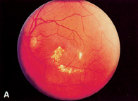
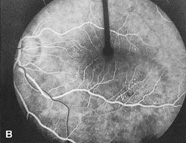
Idiopathic juxtafoveolar retinal telangiectasis is characterized by abnormal retinal capillaries and variable intraretinal exudation after exclusion of an ocular or systemic cause.45,46 Onset is in adulthood, and patients present with blurred central vision. The hallmark lesions are small, perifoveal, and parafoveal telangiectatic capillaries that demonstrate tortuosity, dilation, and irregular caliber and may be associated with lipid exudation in some cases (Fig. 7). Microaneurysms can be present.47 The abnormal vessels are either localized to an area that usually is temporal to the fovea and measures between 1 and 5 mm in diameter or scattered diffusely throughout the macula.45,47 Macular edema or lipid exudation can decrease vision.48 An updated classification by Gass and Blodi in 1993 divides patients based on biomicroscopic and IVFA features into three groups (1, 2, and 3) and subgroups (A and B).49 Overall, the telangiectases appear to be related to retinal capillary leakage in group 1, capillary diffusion abnormalities in group 2, and capillary occlusion in group 3.

Fig. 7. A. Fundus photograph of a patient with idiopathic unilateral juxtafoveal retinal telangiectasis. Notice the localized lipid exudate. B. IVFA reveals focal areas of telangiectasis and leakage.

Group 1A is composed of unilateral congenital parafoveal telangiectasis, typically occurring in young to middle-aged male patients. A small area of easily visible retinal vascular abnormalities is present in the temporal macula. There is mild visual loss from macular edema. Laser photocoagulation to areas of leakage may improve vision, Group 1B consists of middle-aged men with a small area of unilateral idiopathic retinal telangiectasis at the edge of the foveal avascular zone, Laser photocoagulation usually is not performed because of the close proximity to the fovea. Group 1 unilateral juxtafoveal telangiectases represent a localized form of congenital retinal telangiectasis (Coats' syndrome).

In contrast, group 2 consists of bilateral acquired idiopathic parafoveal telangiectases that become clinically apparent later in the fifth and sixth decade.49 The zones of telangiectasis tend to be symmetric and measure up to 1 disc diameter, with a preference for the temporal parafoveal region. Retinal edema and hard exudates are minimal. So called “right angle” veins are seen draining the telangiectatic areas. Underlying retinal pigment epithelial alterations and superficial retinal refractile deposits can develop. Some patients develop a small, yellow, pseudovitelliform lesion within their fovea. Visual loss usually is mild but can be severe. Pericentral scotomas are common. Later in the course, choroidal neovascularization (CNV), which often is subfoveal, foveolar atrophy, and intraretinal pigment plaques may develop.46 Histopathologic study of bilateral juxtafoveal telangiectasis reveals endothelial cell and pericyte degeneration, as well as retinal capillary narrowing associated with endothelial cell basement membrane proliferation.50 Photocoagulation probably is not beneficial because visual loss appears to result from retinal atrophy rather than exudation; however, it may play a role in treating secondary CNV. Surgical removal of subfoveal CNV has had poor results because of its adherence with the overlying neurosensory retina.51

Group 3 is an uncommon variant with bilateral prominent idiopathic parafoveal telangiectasis and minimal exudation. Visual loss occurs from progressive parafoveal capillary occlusion. There is no leakage from the capillary bed. These patients may have related systemic disease.

Retinal vascular telangiectasia also can occur in association with some systemic and ocular conditions. Sickle cell disease and bilateral carotid artery obstruction can mimic the idiopathic version of this condition.52,53 Dilated perifoveal capillaries also have been seen with Stargardt's disease and facioscapulohumeral muscular dystrophy.54,55 Retinal telangiectasia may occur with retinal vein occlusion or radiation retinopathy or represent an atypical presentation of background diabetic retinopathy.56 In one series, 62% of patients with bilateral juxtafoveolar retinal telangiectasis had abnormal glucose metabolism.57 For this reason, all affected patients should undergo a glucose tolerance test and a fasting blood glucose to rule out latent diabetes mellitus.

Back to Top
CONGENITAL RETINAL TELANGIECTASIS (COATS' DISEASE)
In 1908, Coats published his findings on patients with retinal diseases associated with massive subretinal exudation.58 He divided the patients into three groups; (1) those with retinal vascular disease, (2) those without retinal vascular disease, (3) and those with a large arteriovenous communication. It is accepted that Coats' third group were patients with retinal capillary hemangiomas (von Hippel angiomas).59 Coats' first group consisted of patients with the severe form of a disease that Reese has since renamed congenital retinal telangiectasis.60 The term Coats' disease should be reserved for patients with congenital retinal telangiectasis associated with marked hard exudation.

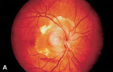
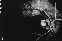
Congenital retinal telangiectasis (Coats' disease) is an idiopathic retinal vascular disorder that usually affects young male patients unilaterally in their first or second decade of life. Congenital retinal telangiectasis, however, can affect patients of either gender and become manifest at any age. Up to one third of patients are older than 30 years of age at the time of presentation.14 There is no defined familial inheritance. Patients may present with decreased vision, as well as strabismus or leukocoria in children. The hallmark feature of congenital retinal telangiectasis is localized fusiform aneurysmal dilations of the retinal vessels reminiscent of tiny light bulbs60 (Fig. 8A). These aneurysms and telangiectatic retinal vessels often are adjacent to areas of retinal capillary nonperfusion with dilated intercapillary spaces. The vascular anomalies can occur anywhere in the fundus and may involve the capillaries, arteries, and veins. Other findings may include vascular loops and beading, retinal neovascularization, hemorrhagic retinal macrocysts, and segmentally dilated capillaries.61 Leakage from the incompetent vasculature may lead to retinal edema, lipid deposition, or, in severe cases, an exudative retinal detachment. The extent of retinal involvement is variable. Infants and children often are more severely affected with extensive vascular involvement and massive subretinal lipid exudate. Vascular leakage may produce a cloudy yellow or green subretinal exudate, which gravitates toward the posterior pole. When the serous component of the exudate resorbs, the yellow exudate remains in the macula and may eventually develop into a fibrovascular macular scar. In contrast, a more localized area of telangiectasis may decompensate in adulthood and present as group 1 idiopathic juxtafoveal retinal telangiectasis. IVFA shows aneursymal dilation and leakage from telangiectatic retinal vessels adjacent to areas of retinal capillary dilation and nonperfusion (see Fig. 8B). The clinical course is variable but often progressive. Secondary complications include cataract, vitreous hemorrhage, neovascular or angle-closure glaucoma, CNV, and phthisis bulbi in severe cases. Histopathologic study reveals a characteristic proteinaceous exudate in the subretinal space containing cholesterol clefts and foamy histiocytes. Intraocular calcification rarely has been demonstrated in Coats' disease, which is an important consideration when differentiating this entity from retino-blastoma.62

Fig. 8. A. Unilateral congenital retinal telangiectasis and massive lipid exudation (Coats' disease) in a 16-year-old boy. Notice the characteristic telangiectatic vessels resembling light bulbs. B. IVFA of the same area revealed hyperfluorescence of the telangiectasia and areas of capillary nonperfusion.

The syndrome of Leber's miliary aneurysms is a form of congenital retinal telangiectasis that is intermediate in severity. Unilateral, congenital retinal telangiectasis, Leber's miliary aneurysms, and idiopathic juxtafoveal retinal telangiectasis represent the clinical spectrum of a single primary retinal vascular disorder.63

Treatment of congenital retinal telangiectasis is directed to close the leaking telangiectatic vessels to permit resorption of exudate. The treatment modality depends on the location and extent of the lesions.64 Laser photocoagulation is the treatment of choice for small telangiectatic lesions. Cryotherapy may be required for extensive peripheral lesions or for lesions associated with exudative retinal detachments. Multiple treatment sessions may be required. Treatment is directed at the leaking telangiectasis and adjacent zones of capillary nonperfusion. External drainage of subretinal fluid with or without scleral buckling can be used if there is a severe exudative retinal detachment.65 Vitrectomy occasionally is indicated for severe or tractional retinal detachments.66 New telangiectasis may appear in areas of retina that were previously uninvolved; therefore, close follow-up is imperative. Despite successful closure of telangiectasis, visual results may be poor, especially when there is macular exudate at presentation.64 Coats' syndrome or a “Coats-like response” (retinal detachment with massive subretinal exudation) has been associated with other ocular and systemic diseases, including retinopathy of prematurity, branch retinal vein obstruction, retinitis pigmentosa, pars planitis, and muscular dystrophy and hemifacial atrophy.49,67–71

Back to Top
CAPILLARY HEMANGIOMA OF THE RETINA
The capillary hemangioma of the retina (also known as angiomatosis retinae or von Hippel's disease) was described as a clinical entity by Eugen von Hippel in 1904.72 The retinal lesions are hamartomas, therefore cytologically benign, and can be inherited in an autosomal dominant fashion with variable penetrance.73 A sporadic, nonheritable form of the disease probably also exists. There is no gender preference. These lesions may be more common in whites.33

The association between these retinal vascular tumors and hemangiomatous cysts of the cerebellum was first recognized in 1926 by Lindau, and this association is referred to as von Hippel-Lindau disease.74 von Hippel-Lindau disease is defined as the combination of one or more capillary hemangiomas of the retina in association with a cerebellar hemangioblastoma. This is an autosomal dominant phakomatosis with variable penetrance.43 Many cases occur as a spontaneous mutation, and thus the family history is positive in only approximately 20% of affected persons.43 Lindau estimated that 25% of all patients with retinal lesions have or develop an intracranial mass.74 Other systemic findings associated with von Hippel-Lindau syndrome include pheochromocytoma, renal cell carcinoma (which is the most common cause of death in these patients), and ovarian, pancreatic, and epididymal cysts.43,75,76 Retinal capillary hemangioma is the first manifestation in approximately 50% of these patients.77 When von Hippel-Lindau disease is suspected, systemic workup should include either a MRI or CT scan of the brain with attention to the posterior fossa. An abdominal CT scan and laboratory analysis for the presence of a pheochromocytoma also should be performed.75 When the family history is positive, affected patients require lifelong medical evaluations by an internist, since the protean systemic manifestations can develop years after the retinal tumors are noted.77 The gene for von Hippel-Lindau disease has been localized at 3p25-26, and genetic testing is available for affected patients.78 Retinal capillary hemangiomas also have been reported in neurofibromatosis and Marshall-Stickler syndrome.79,80

Clinically, retinal capillary hemangiomas are smooth, oval, pink or red intraretinal masses (Fig. 9A). Gliosis and retinal pigmentary changes can occur on the lesion surface to alter this appearance.75 Retinal hemorrhage and exudate often surround the tumors. When fully developed, retinal capillary hemangiomas are fed by markedly dilated, often beaded, and tortuous arteries and are drained by similarly appearing veins (see Fig. 9B). When the lesions are small, however, the feeding and draining vessels may not appear abnormal.73 The tumors are typically between 1 and 5 mm, although larger lesions measuring 10 mm have been reported.75 The lesions can occur anywhere in the fundus but are most frequently seen in the temporal equatorial region and on the optic nerve73,75 (Fig. 10A). They are multiple in approximately one third of affected eyes and bilateral in approximately one half of affected persons.73,75,81 Growth of preexisting lesions, as well as development of new retinal capillary hemangiomas in previously uninvolved retina, has been documented. Unusual retinal vascular hamartomas other than retinal angiomas have been detected in patients with von Hippel-Lindau disease and their relatives.82 IVFA demonstrates rapid dye transit through the tumor with profuse leakage of fluorescein during the recirculation phase (see Fig. 10B). Pathologic study of the retinal capillary hemangioma lesions demonstrates thin-walled capillaries surrounded by stromal cells.83 The blood-filled spaces tend to be small.81

Fig. 9. A. Small retinal capillary hemangioma in a patient with von Hippel-Lindau disease. There is a distinct feeding arteriole and draining venule as well as a small amount of lipid exudation. IVFA revealed rapid filling of the tumor. B. larger retinal capillary hemangioma with prominent feeding.

Fig. 10. A. Retinal capillary hemangioma of the optic disc (endophytic type) associated with retinal edema and exudate. B. IVFA shows early hyperfluorescence of the lesion in the arterial phase, (B, courtesy of James J, Augsburger, MD)

The usual age of presentation for a patient with a retinal capillary hemangioma is the second or third decade of life. On occasion, asymptomatic lesions are noted on routine examination. Loss of vision usually occurs when the capillaries associated with the tumor become incompetent and exudate extends into the macula. Even small, peripheral retinal capillary hemangiomas can produce remote exudation into the fovea.75 If these hemangiomas are not treated, total exudative retinal detachment can ensue with secondary iris neovascularization and neo-vascular glaucoma. Epiretinal membranes, vitreous hemorrhage, retinal neovascularization, and both fractional and rhegmatogenous retinal detachments also can occur.84,85

The clinical appearance of retinal capillary hemangiomas located at the optic nerve varies considerably from that of capillary hemangiomas of the retinal periphery. The optic nerve lesions have been described in three varieties.86 The most easily recognized type is the endophytic, which grows anteriorly into the vitreous cavity. It is similar in color and appearance to the peripheral retinal lesions while lacking abnormally dilated feeding and draining vessels (see Fig. 10A). The sessile and exophytic types grow toward the subretinal space and also lack large feeding and draining retinal vessels. The latter two varieties can be difficult to differentiate from peripapillary CNV, choroiditis, choroidal hemangiomas, or optic nerve edema. Stereoscopic IVFA may help to differentiate these entities.86 Clinically, sessile hemangiomas appear as flat, ill-defined, yellow-orange lesions visible underneath grayish, thickened overlying retina. The endophytic type is similar in appearance but has a nodular growth pattern. Visual loss occurs secondary to serous or exudative fluid that extends into the macula, often with hard exudate, Rarely does the serous retinal detachment become as severe as with peripheral retinal capillary hemangiomas. Optic disc and retinal capillary hemangiomas can occur in the same eye together, or a patient can have a disc hemangioma in one eye with a retinal hemangioma in the other. Bilateral optic disc capillary hemangiomas have been described.87

Retinal capillary hemangioma can simulate congenital retinal telangiectasis with exudative detachment (Coats' disease). The lack of either “light bulb” telangiectasis or peripheral retinal capillary nonperfusion on IVFA, coupled with the presence of feeding and draining vessels leading to a retinal mass, usually reveals the proper diagnosis. Other entities in the differential diagnosis are grade III arteriovenous communications and cavernous hemangiomas of the retina.

Based on the natural history of progressive visual loss and their potential for growth, most authorities recommend some type of obliterative therapy, even when the visual acuity is unaffected. The treatment modality depends on the size, location, and number of lesions. Retinal capillary hemangiomas can be treated with laser photocoagulation or cryotherapy, diathermy, or radiotherapy.73,88–92 Techniques for laser photocoagulation include direct treatment of the lesion, laser to the feeding arteriole to reduce tumor blood flow before direct treatment, and scatter treatment around the lesion to prevent posttreatment extension of exudate. Radiotherapy is a newer treatment for this entity. Plaque radiotherapy appears useful for lesions with extensive exudative detachments, whereas charged particle beam irradiation may be used for epipapillary lesions.92 Peeling of epiretinal macular membranes has been reported after obliteration of peripheral lesions.84 On occasion, obliterative treatment may lead to total exudative retinal detachment (ablatio fugax) or vitreous hemorrhage. Vitrectomy may be indicated for severe exudative or fractional retinal detachment.73,85,93 Eye wall resection has been attempted to treat exceptionally large tumors.94 Despite treatment, the prognosis for visual acuity with larger lesions remains guarded.75,95 In contrast, retinal capillary hemangiomas of the optic disc usually are not treated unless they become symptomatic. Obliterative treatment strong enough to destroy the tumor at the optic nerve can result in total loss of vision.

Back to Top
VASOPROLIFERATIVE TUMORS OF THE FUNDUS
Shields and associates describe 12 patients with fundus lesions that resembled retinal capillary hemangiomas on clinical examination but were believed to develop on an acquired basis.96 These lesions were characterized as a vascularized mass of the sensory retina with a yellow-red appearance but lacked the enlarged, tortuous feeding and draining vessels typically seen with von Hippel-Lindau retinal capillary hemangiomas. Also, there was no association found with von Hippel-Lindau disease in these individuals, although 50% had known systemic hypertension. The term presumed acquired retinal hemangiomas initially was coined to describe this entity. Campochiaro and Conway report five additional patients with similar lesions.97 Other authors have used terms such as hemangioma-like, angioma, angiomatous mass, and acquired peripheral retinal telangiectasis to describe this entity.98–100

In 1995, Shields and associates characterized 103 patients with these acquired retinal hemangiomas and recommended using the term vasoproliferative retinal tumors to describe these lesions.101,102 The lesions can be unilateral or bilateral and are characterized as either idiopathic (primary) or secondary to preexisting ocular disease. Associated preexisting ocular disorders include intermediate uveitis, retinitis pigmentosa, toxoplasmosis, toxocariasis, his-toplasmosis, retinochoroidal coloboma, traumatic chorioretinopathy, familial exudative vitreoretinopathy, Coats' disease, status postretinal detachment repair, and cryotherapy.100,102 Lesions usually are solitary but can be multiple or diffusely involve the eye and have a propensity to occur in the inferior peripheral retina. Associated ocular findings that may impair vision include intraretinal exudation, exudative retinal detachment, vitreous cells, vitreous hemorrhage, retinal pigment epithelial hyperplasia, premacular fibrosis, and macular edema. The macular changes may be remote from the vasoproliferative tumor. IVFA reveals rapid filling of the vasoproliferative tumor through an undilated or minimally dilated retinal arteriole in the arterial phase and staining of the lesion in the late phases.102 Ultrasonography reveals medium to high internal reflectivity and acoustic solidity without choroidal excavation.102 The pathogenesis of vasoproliferative retinal tumors is not clear. It has been hypothesized that these lesions represent exuberant vascularization of the retina or retinal pigment epithelium or reactive gliosis.101,102 Histopathologic study in one case demonstrated retinal gliosis, dilated ectatic retinal vessels, and a preretinal neovascular membrane.103 Management of these lesions has included observation, cryotherapy, laser photocoagulation, and plaque radiotherapy, depending on the size and location of the lesion and the associated complications.97,102,104

Back to Top
CAVERNOUS HEMANGIOMA OF THE RETINA
Cavernous hemangioma of the retina is a rare benign vascular hamartoma that can arise anywhere in the fundus or from the optic nerve. Reese initially described this clinical entity, although Gass is credited as definitively identifying retinal cavernous hemangiomas as distinct from von Hippel lesions or congenital retinal telangiectasis.17,105 The cavernous hemangioma of the retina usually is sporadic, although occasional familial cases have been reported, and it may be more common in women.14 These lesions are congenital but usually are not detected until the second decade.

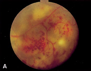
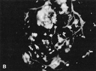
Ophthalmoscopically, they have a distinctive appearance. Cavernous hemangiomas of the retina consist of multiple, elevated saccular lesions arising from the inner retina, giving the appearance of a cluster of grapes (Fig. 11A and B). Each saccule measures from 100 μm to as large as 1500 μm, with the entire lesion typically measuring between 2 and 6 mm.75 The saccules typically appear dark red and are adjacent to a retinal vein. Individual aneurysms may occur separate from the main lesion. Lipid exudation rarely occurs, although associated gliosis may impart a whitish, fibrous, plaque-like-appearance to its surface.105 Hemorrhage on the surface of the lesion has been noted, and vitreous hemorrhage also can occur.105 Growth of these lesions is rare.73,106 Unlike the retinal capillary hemangioma of von Hippel, the surrounding retinal vessels are of normal caliber.107,108 On IVFA, the lesions usually fill slowly, and late leakage of dye is uncommon. A plasma erythrocyte level often is seen within some of the sacculi on IVFA (see Fig. 11C). Visual loss is uncommon, but when it is present, it usually occurs secondary to vitreous hemorrhage. Epiretinal membrane formation, foveal dragging, and am-blyopia also have been reported. Treatment of these lesions is rarely indicated. On occasion, cryotherapy, laser photocoagulation, or vitrectomy may be performed for recurrent visually disabling vitreous hemorrhage.106 Histopathologic study shows large, blood-filled vascular spaces in the inner retina (Fig. 12).

Fig. 11. A. Cavernous hemangioma of the retina. Notice the overlying fibroglial proliferation and satellite lesions on the optic disc. B. A plasma (fluorescein)-erythrocyte level is noted within some of the sacculi on IVFA. C. Cavernous hemangioma of the retina may be located adjacent to or on the optic nerve.

Fig. 12. Histopathologic study of a retinal cavernous hemangioma of the optic disc. Notice multiple, small, thin-walled, blood-filled spaces. (Courtesy of the Armed Forces Institute of Pathology, Washington, DC)

Cavernous hemangioma of the retina has been implicated as a part of an oculoneural cutaneous syndrome.109,110 Associated lesions include hemangiomas of the skin, especially on the back of the neck, as well as seizures, presumably from hemangiomas of the brain. The brain hemangiomas usually occur in the midbrain or cerebral cortex and can be difficult to detect with arteriography or CT scanning.111 Individuals with this syndrome are more likely to have multifocal or bilateral retinal cavernous hemangiomas,109

Back to Top
CHOROIDAL HEMANGIOMA
Choroidal hemangiomas are congenital hamartomatous vascular tumors that occur in two clinically and histopathologically distinct varieties: circumscribed and diffuse.

Circumscribed choroidal hemangiomas are discrete, smooth-surfaced tumors and are characteristically round or oblong33,112 (Fig. 13A). They usually measure between 2 and 6 mm in diameter, although up to 17 mm diameter has been reported.109 Thickness varies from 1 to 6 mm. They usually are found in the macular area or peripapillary region, and the posterior tumor margin typically is located within two disc diameters from the fovea or optic nerve.113,114 Their coloration is a characteristic reddish orange. This distinct color allows them to be differentiated from other amelanotic choroidal tumors. When flat, choroidal hemangiomas can be difficult to differentiate from the background choroidal pattern. Although they are vascular, choroidal hemangiomas rarely show large, distinct, inherent tumor vessels on ophthalmoscopic examination. Retinal pigment epithelial alterations frequently occur over the surface of these lesions. Fibrous metaplasia of the retinal pigment epithelium commonly occurs with time and results in a whitish plaque-like surface. Cystoid degeneration of the overlying retina or even bullous retinoschisis also, can develop.112 Retinal neovascularization and CNV have been described.115,116

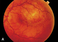
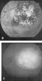
Fig. 13. A. Circumscribed choroidal hemangioma with fibrous metaplasia on its surface. B. IVFA during arterial phase. Notice the large, early filling of choroidal spaces within the tumor, producing a mottled appearance. C. ICG shows diffuse hyperfluorescence of the circumscribed choroidal hemangioma. This is followed by wash out of ICG dye in late phase angiogram.

Decreased vision secondary to associated serous subretinal fluid is the usual reason affected patients seek medical attention. This decreased vision most commonly occurs in the third to fifth decade of life. Total exudative retinal detachment with shifting fluid can develop. Circumscribed choroidal hemangiomas occasionally enlarge slightly over prolonged observation.117 IVFA shows early filling of the hemangioma concomitant with the choroidal circulation, although this pattern is variable and nondiagnostic33 (see Fig. 13B). ICG angiography is more useful than IVFA to define circumscribed choroidal hemangiomas. ICG dye fills the intralesional vessels early. There is intense ICG hyperfluorescence in the mid-angiogram (see Fig. 13C), followed by late clearing of the dye with a “washout” phenomenon. 118,119 Ultrasonography reveals a placoid choroidal mass with acoustic solidity and high internal reflectivity secondary to multiple interfaces within the tumor.

In the past, many eyes containing circumscribed choroidal hemangiomas were enucleated because the lesions were confused with amelanotic choroidal melanomas. Careful ophthalmoscopic examination, with attention to the lesion's color and the presence of inherent uveal pigmentation or large, distinct choroidal vessels, usually provides the correct diagnosis. Other lesions in the differential diagnosis include choroidal granuloma, metastatic tumor, and choroidal osteoma. Angiography (IVFA, ICG) and ultrasonography are helpful in differentiating these entities. Circumscribed choroidal hemangiomas are not inherited and are not associated with systemic abnormalities.

Diffuse choroidal hemangiomas are larger and flatter than the circumscribed variety, with more indistinct borders.33 This lesion resembles diffuse choroidal thickening, but some nodular thickening can occur in the posterior pole. Diffuse choroidal hemangiomas commonly cover over one half of the fundus, extending into the equatorial zone. The lesion imparts a bright red reflex to the fundus, resulting in the so-called “tomato catsup” fundus.33 Comparison of this red reflex with a normal opposite eye can be helpful in confirming the diagnosis. On ophthalmoscopic examination, the normal underlying choroidal pattern is obscured. The overlying retinal vessels also may be affected. Dilation and tortuosity of the retinal vessels, as well as peripheral arteriovenous communications, have been noted.112 Exudative retinal detachment can occur. Ultrasound of the diffuse variety demonstrates generalized choroidal thickening and optic nerve cupping. Both MRI and CT scan can show diffuse choroidal hemangioma.120

Diffuse choroidal hemangiomas are associated with Sturge-Weber syndrome (encephalotrigeminal angiomatosis), a noninherited phakomatosis, or a variant such as Klippel-Trenaunay-Weber or Milles' syndrome.43,112,113,121 The Sturge-Weber triad consists of a facial nevus flammeus (a large vascular telangiectasia, not a true hemangioma), ipsilateral buphthalmos, and contralateral seizures caused by ipsilateral leptomeningeal hemangiomatosis. The diffuse choroidal hemangioma is not a requisite part of the syndrome. It is present in approximately 50% of affected persons and can be bilateral.122 The associated glaucoma can be infantile, juvenile, or neo-vascular. When a person has an isolated nevus flammeus without the full Sturge-Weber syndrome, the probability of having an accompanying diffuse choroidal hemangioma is about 2%.33 Like the circumscribed variety, diffuse choroidal hemangioma can lead to exudative retinal detachment, secondary retinal and iris neovascularization, and neovascular glaucoma.

Pathologic study of choroidal hemangiomas shows a distinct tumor with dilated, blood-filled spaces lined by endothelium.113 Often, the overlying retinal pigment epithelium reveals hyperplasia, metaplasia, or even ossification. The overlying retina can demonstrate outer plexiform cystic changes. Most circumscribed choroidal hemangiomas are either of the pure cavernous type or the mixed cavernous and capillary type. The diffuse tumors usually are composed of both capillary and cavernous components.33

When symptomatic, circumscribed choroidal hemangiomas can be treated with obliterative therapy. Both light scatter argon laser photocoagulation to the tumor's surface and intense obliteration with either argon or krypton laser or xenon photocoagulation have been advocated.111 Multiple treatments may be required because of reaccumulation of sub-retinal fluid. Diode laser hyperthermia without photocoagulation is being investigated as an alternative treatment.123 Cryotherapy and penetrating diathermy are of limited use because of the posterior location of the tumors. Radiation therapy has been used with success in eyes with thick choroidal hemangiomas and bullous nonrhegmatogenous retinal detachments, and eyes that have not responded to other modalities.124,125 External beam irradiation and plaque and proton beam radiotherapy have been used. On occasion, external drainage of subretinal fluid with or without scleral buckling in conjunction with laser photocoagulation has been used to treat diffuse choroidal hemangiomas associated with large exudative retinal detachments.33 Alternatively, vitrectomy, endolaser, and internal drainage of subretinal fluid can be performed. The visual prognosis remains guarded because of significant secondary retinal changes that can occur over the tumor's surface and the lesion's propensity for recurrent leakage.

Back to Top
IDIOPATHIC POLYPOIDAL CHOROIDAL VASCULOPATHY
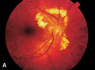
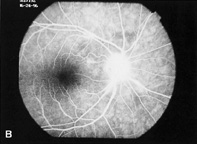
Idiopathic polypoidal choroidal vasculopathy (IPCV), also known as posterior uveal bleeding syndrome or multiple recurrent serosanguineous retinal pigment epithelial detachment syndrome, is characterized by choroidal vascular anomalies.126–128 It is more common in middle-aged women and is associated with African-American race and systemic hypertension.129 The lesions in IPCV are unilateral or bilateral orange-red subretinal polypoidal saccular dilations arising from an inner choroidal vascular network130 (Fig. 14A). The polypoidal lesions typically originate from the peripapillary choroidal circulation but also have been described in the macular region and in the peripheral fundus.131 Visual loss may result from subretinal hemorrhage, vitreous hemorrhage, or subretinal exudate from the polypoidal lesions. IVFA angiography reveals hyperfluorescence and pooling of fluorescein in the polypoidal choroidal lesions. ICG angiography characteristically reveals a dilated network of inner choroidal vessels, which terminate in multiple saccular dilations that correspond clinically to the orange subretinal lesions130,132 (see Fig. 14B).

Fig. 14. A. Small orange-red subretinal lesions in idiopathic polypoidal choroidal vasculopathy are partially obscured by adjacent subretinal hemorrhage. B. ICG hyperfluorescence demonstrates the polypoidal choroidal vascular network, which is contrasted with the hypofluoerscent subretinal hemorrage. (A, courtesy of James J. Augsburger, MD)

Differentiation of IPCV from CNV secondary to age-related macular degeneration is important, since the treatment and prognosis of these entities differs. IPCV is differentiated from age-related macular degeneration by the absence of drusen and subretinal fibrosis, as well as its characteristic clinical and ICG features. The optimal management of IPCV is not clear. Lesions with hemorrhagic complications can present acutely with visual loss but occasionally resolve spontaneously with visual recovery. Laser photocoagulation of the polypoidal lesions may be considered if subretinal fluid, exudate, or hemorrhage threatens or involves the fovea.130

Back to Top
IDIOPATHIC RETINAL VASCULITIS, ANEURYSMS AND NEURORETINITIS
Idiopathic retinal vasculitis, aneurysms, and neuroretinitis (IRVAN) is an uncommon syndrome characterized by retinal vasculitis, multiple macro-aneurysms, neuroretinitis, and peripheral retinal capillary nonperfusion.133 This initially was described in detail by Schatz and Kincaid in two patients with bilateral retinal arteritis, multiple macro-aneurysms, neuroretinitis, and uveitis.134 Thirteen additional patients have been reported with this disorder,133,135–137 It is not clear whether this syndrome is a primary vascular or inflammatory disorder, but it is included in this chapter based on its unusual retinovascular features.

This syndrome typically affects young healthy individuals with a female preponderance. Despite the significant retinal findings, patients often are asymptomatic at presentation. In the largest series of 10 patients, all three features described by the acronym IRVAN were required for diagnosis.133 This includes the presence of numerous aneurysmal dilations of the optic nerve head and retinal arterioles at or near their major branching sites. The aneurysms measure between 75 and 300 μm in diameter and may have a triangular, Y-shaped or coiled configuration. Aneurysmal changes at the optic nerve head may be associated with ectatic changes and apparent elongation of the adjacent arteries (Fig. 15A). The vascular abnormalities in IRVAN have a propensity to leak, leading to exudative retinopathy, but do not have a predisposition to hemorrhage. Subretinal, intraretinal, or vitreous hemorrhage secondary to aneurysms has not been associated with IRVAN. This is contrasted with senile acquired macroaneurysms, which occur in older individuals with systemic hypertension, are round, and may be associated with either hemorrhage or serous exudation. IVFA is useful to delineate aneurysmal changes in IRVAN. Vasculitis is demonstrated by staining of the arterial and venous walls on IVFA, and the arterioles appear to be more severely affected (see Fig. 15B). Neuroretinitis is manifested clinically as optic nerve head swelling and as late diffuse staining of the optic nerve head on IVFA. Extensive peripheral retinal capillary nonperfusion with sequelae of retinal ischemia, such as neovascularization of the retina or anterior segment, may occur. Vitreous hemorrhage may occur secondary to retinal neovascularization. Vitritis has been described in IRVAN and may be accompanied by anterior uveitis. IRVAN is differentiated from Bales' disease, which has a predilection for young male patients, an association with tuberculin hypersensitivity, more marked uveitis, retinal venous inflammation, and a lack of multiple macroaneurysms.133 Although some cases of IRVAN maintain good visual acuity, severe visual loss may result from exudative maculopathy or sequelae of retinal ischemia.

Fig. 15. A. Fundus photograph of IRVAN demonstrates aneurysms and apparent vascular elongation at the optic nerve with secondary exudation, B. JVFA of IRVAN reveals late staining of the optic nerve head and vasculitis. (A, courtesy of Carmen A, Puliafito, MD)

Extensive medical investigations have not revealed any systemic associations. Systemic corticosteroids do not appear to have a beneficial effect on uveitis or retinal ischemia. There is limited experience regarding laser photocoagulation to aneurysms to treat eyes with exudative retinopathy threatening the fovea. Branch artery occlusion was described in one case after laser photocoagulation to a single aneurysm.133 Chang and colleagues recommend observing eyes without retinal neovascularization and performing panretinal laser photocoagulation for eyes that have developed retinal neovascularization or have signs of rapidly progressive anterior segment ischemia.133 Vitrectomy has been performed for eyes with persistent vitreous hemorrhage.

Back to Top
VARIX OF THE VORTEX VEIN
A varix of the vortex vein represents a large dilation of the vortex vein ampulla that may be noted during indirect ophthalmoscopic examination. It typically becomes more prominent when the eye is directed in eccentric gaze, and it often is located at the equator in the nasal retina.138 This entity is asymptomatic and may be overlooked during examination. On occasion, a varix of the vortex vein may be large enough to simulate a solid choroidal lesion such as a small choroidal melanoma.138–140 However, the clinical features and dynamic nature of a vortex vein varix are characteristic for this entity.

The varix appears as a dark red, smooth, dome-shaped elevation located deep to the retina. It can measure between 3 and 6 mm in diameter and up to 2.5 mm in thickness.138 It can be unilateral or bilateral, and more than one varix may be noted within one eye.138,141 It is typically reported in middle-aged to elderly patients, although it has been noted in patients as young as 23 years,142 There are no known systemic associations. The varix can compress the adjacent choroid, which increases visibility of choroidal pigment at the edge of the ampulla. This feature may lead to diagnostic confusion with choroidal melanoma. The continuity of the lesion with the choroidal vasculature and the fluctuating nature of the varix are helpful to confirm its diagnosis. The varix becomes more prominent with positioning of the affected eye in the direction of the varix (Fig. 16A). Also, the varix may become engorged by the Valsalva maneuver or by lowering the individuals head below the level of the heart.141 Positioning the eye in primary position, elevating the head, or applying pressure on the globe digitally or with a contact lens can make the varix less prominent or cause it to disappear140 (see Fig. 16B). The pathogenesis of a varix of the vortex vein has been attributed to gaze-evoked kinking of the extrascleral portion of the vortex vein, gaze-evoked narrowing of the scleral emissary canal, and increased ocular venous pressure.139,141,143

Fig. 16. A. Gaze-evoked prominence of a vortex vein varix at the inferonasal equator. B. The vortex vein varix disappears in primary gaze. C. ICG angiography shows early filling of the vortex vein varix at 40 seconds. D. ICG hyperfluorescence of the vortex vein varix is decreased in primary gaze. (A to D courtesy of Jerry A. Shields, MD, and Carol L. Shields, MD)

Diagnostic tests include B-scan ultrasound, IVFA, ICG angiography, and color Doppler imaging. B-scan ultrasound reveals acoustic solidity and gaze-evoked enlargement of the lesion. Increased intraocular pressure induced by the ultrasound probe may cause the lesion to disappear.139 Low to medium internal reflectivity is noted on Ascan. IVFA shows mild patchy choroidal fluorescence of the vortex vein ampulla in the venous phase with late pooling of dye in the outer choroid. ICG angiography reveals early hyperfluorescence with late pooling of dye in the varix, and gaze-evoked fluctuation of ICG hyperfluorescence may be noted143 (see Fig. 16C and D). Color Doppler imaging shows a vascular lesion of venous origin that fills when the eye is directed to the quadrant of the lesion.144

Back to Top
REFERENCES

1. Duke-Elder S, Wybar KC: System of ophthalmology. In Duke-Elder S (ed): The Anatomy of the Visual System, vol 2, pp 356–358. St Louis: CV Mosby, 1961

2. Awan KJ: Familial chorioretinal vascular anastomoses and congenital cataracts. J Pediatr Ophthalmol Strab 17:384, 1980

3. Ohno-Matsui K, Morishima N, Ito M et al: Posterior routes of choroidal blood outflow in high myopia. Retina 16:419, 1996

4. Lichter PR, Schmickel RD: Posterior vortex vein and congenital glaucoma in a patient with trisomy 13 syndrome. Am J Ophthalmol 80:939, 1975

5. Mann IC: Developmental Abnormalities of the Eye, pp 138–139. Philadelphia: JB Lippincott, 1957

6. Brown GC, Tasman WS: Congenital Anomalies of the Optic Disc, pp 31–65. New York: Grune & Stratton, 1984

7. Duke-Elder S: System of Ophthalmology, ed 2, vol 3, pp 786–787. St Louis: CV Mosby, 1963

8. Birnholz JC, Farrell EE: Fetal hyaloid artery; Timing of regression with US. Radiology 166:781, 1988

9. Yap EY, Buettner H: Traumatic rupture of a persistent hyaloid artery. Am J Ophthalmol 114:225, 1992

10. Goncalves A, Cruysberg JR, Draaijer RW et al: Vitreous haemorrhage and other ocular complications of a persistent hyaloid artery. Doc Ophthalmol 92:55, 1996

11. Chen TL, Yarng SS: Vitreous hemorrhage from a persistent hyaloid artery. Retina 13:148, 1993

12. Eller AW, Jabbour NM, Hirose T, Schepens CL: Retinopathy of prematurity: The association of a persistent hyaloid artery. Ophthalmology 94:444, 1987

13. Liebrich R: Demonstrations of diseases of the eye: A persistent hyaloid artery and vein. Trans Pathol Soc London 22:221, 1871

14. Gass JDM: Stereoscopic Atlas of Macular Diseases; Diagnosis and Treatment, vol 1, pp 208–217, 438–444, 494–502, 844–860, 868–870, St Louis: CV Mosby, 1997

15. Degenhart W, Brown GC, Augsburger JJ et al: Prepapillary vascular loops: A clinical and fluorescein angiographic study. Ophthalmology 88:1126, 1981

16. Shakin EP, Shields JA, Augsburger JJ, Brown GC: Clinico-pathologic correlation of a prepapillary vascular loop, Retina 8:55, 1988

17. Brucker AJ, Michels RG, Fine SL: Congenital arterial loops and retinal arterial occlusion. Am J Ophthalmol 84:220, 1977

18. Brown GC, Margargal LE, Augsburger JJ et al: Preretinal arterial loops and retinal arterial occlusion, Am J Ophthalmol 87:646, 1979

19. Strassman IB, Desai UR: Prepapillary vascular loop and a recurrent vitreous hemorrhage. Retina 17:166, 1997

20. Soltau JB, Oik RJ, Gordon JM: Prepapillary arterial loop associated with vitreous hemorrhage and venous retinal macrovessel. Retina 16:74, 1996

21. Grossniklaus H, Thall E, Annable W: Familial prepapillary vascular loops, Arch Ophthalmol 104:1755, 1986

22. Lambert HM, Sipperley JO, Shacklett DE: Autosomal dominant preretinal vascular loops, Retina 3:258, 1983

23. Goldberg MF, Pollack IP, Green WR: Familial retinal arte-riolar tortuosity with retinal hemorrhage. Am J Ophthalmol 73:183, 1972

24. Bartlett WJ, Price J: Familial retinal arterial tortuosity with retinal hemorrhage. Am J Ophthalmol 95:556, 1983

25. Wells CG, Kalina RE: Progressive inherited retinal arterio-lar tortuosity with spontaneous retinal hemorrhages. Ophthalmology 92:1015, 1985

26. Sears J, Oilman J, Sternberg P Jr: Inherited retinal arteriolar tortuosity with retinal hemorrhages. Arch Ophthalmol 116:1185, 1998

27. Connelly BW, Gibson GG: The retinal arterioles in coarcta-tion of the aorta: 14 years' observation of a case. Am J Ophthalmol 32:1513, 1949

28. Meredith TA: Inherited retinal venous beading, Arch Ophthalmol 105:949, 1987

29. Stewart MW, Gitter KA: Inherited retinal venous beading, Am J Ophthalmol 106:675, 1988

30. Brown GC, Donoso LA, Margargal LE et al: Congenital retinal macrovessels, Arch Ophthalmol 100:1430, 1982

31. Mansour AM, Walsh JB, Henkind P: Arteriovenous anastomoses of the retina. Ophthalmology 94:35, 1987

32. Archer DB, Deutman A, Krill AE: Arteriovenous communications of the retina, Am J Ophthalmol 75:224, 1973

33. Shields JA, Shields CL: Intraocular Tumors: A Text and Atlas, pp 394–418, 514–535, Philadelphia: WB Saunders, 1992

34. Augsburger JJ, Goldberg RE, Shields JA et al: Changing appearance of retinal arteriovenous malformation. Graefes Arch Clin Exp Ophthalmol 215:65, 1980

35. Pauleikhoff D, Wessing A: Arteriovenous communications of the retina during a 17-year follow-up, Retina 11:433, 1991

36. Mansour AM, Wells CG, Jampol LM, Kalina RE: Ocular complications of arteriovenous communications of the retina. Arch Ophthalmol 107:232, 1989

37. Tilanus MD, Hoyng C, Deutman AF et al: Congenital arteriovenous communications and the development of two types of leaking retinal macroaneurysms, Am J Ophthalmol 112:31, 1991

38. Munoz FJ, Rebolleda G, Cores FJ, Bertrand J: Congenital retinal arteriovenous communication associated with a full-thickness macular hole, Acta Ophthalmologica 69:117, 1991

39. Schatz H, Chang LF, Ober RR et al: Central retinal vein occlusion associated with retinal arteriovenous malformation. Ophthalmology 100:24, 1993

40. Shah GK, Shields JA, Lanning RC: Branch retinal vein obstruction secondary to retinal arteriovenous communication. Am J Ophthalmol 126:446, 1998

41. Boiling JP, Buettner H: Acquired retinal arteriovenous communications in occlusive disease of the carotid artery. Ophthalmology 97:1148, 1990

42. Wyburn-Mason R: Arteriovenous aneurysm of midbrain and retina, facial naevi and mental changes. Brain 66:163, 1943

43. Font RL, Ferry AP: The phakomatoses, Int Ophthalmol Clin 12:1, 1972

44. Bech K, Jensen OA: On the frequency of co-existing racemose haemangiomata of the retina and brain, Acta Psychi-atr Scand 36:47, 1961

45. Horn G, Rabb MF, Lewickey AO: Retinal telangiectasis of the macula: A review and differential diagnosis, Int Ophthalmol Clin 21:139, 1981

46. Gass JDM, Oyakawa RT: Idiopathic juxtafoveal retinal telangiectasis. Arch Ophthalmol 100:769, 1982

47. Mansour AM, Schachat A: Foveal avascular zone in idiopathic juxtafoveolar telangiectasia. Ophthalmologica 207:9, 1993

48. Chopdar A: Retinal telangiectasis in adults: Fluorescein angiographic findings and treatment by argon laser. Br J Ophthalmol 62:243, 1978

49. Gass JD, Blodi B A: Idiopathic juxtafoveolar retinal telangiectasis: Update of classification and follow-up study. Ophthalmology 100:1536, 1993

50. Green WR, Quigley HA, De La Cruz Z et al: Parafoveal retinal telangiectasis: Light and electron microscopy studies, Trans Ophthalmol Soc UK 100:162, 1980

51. Berger AS, McCuen BW, Brown GC, Brownlow RL Jr: Surgical removal of subfoveal neovascularization in idio-pathic juxtafoveolar retinal telangiectasis, Retina 17:94, 1997

52. Asdourian G, Nagpal K, Busse B et al: Macular and perima-cular vascular remodeling in sickling hemoglobinopathies. Br J Ophthalmol 60:431, 1976

53. Campo RV, Reeser FH: Retinal telangiectasia secondary to bilateral carotid artery occlusion. Arch Ophthalmol 101:1211, 1983

54. Deutman AF, Pinckers AJ, Aan de Kerk A: Dominantly inherited cystoid macular edema, Am J Ophthalmol 82: 540, 1976

55. Gurwin EB, Fitzsimons RB, Sehmi KS et al: Retinal telangiectasis in fascioscapulohumeral muscular dystrophy with deafness. Arch Ophthalmol 102:1695, 1985

56. Chew EY, Murphy RD, Newsome DA et al: Parafoveal telangiectasis and diabetic retinopathy, Arch Ophthalmol 104:71, 1986

57. Milay RH, Klein ML, Handelman IL, Watzke RC: Abnormal glucose metabolism and parafoveal telangiectasia, Am J Ophthalmol 102:363, 1986

58. Coats G: Forms of retinal disease with massive exudation, R Lond Ophthalmol Hosp Rep 17:440, 1908

59. Henkind P, Morgan G: Peripheral retinal angioma with exudative retinopathy in adults (Coat's lesion). Br J Ophthalmol 50:2, 1966

60. Reese AB: Telangiectasis of the retina and Coats' disease, Am J Ophthalmol 42:1, 1956

61. Goel SD, Augsburger JJ: Hemorrhagic retinal macrocysts in advanced Coats' disease. Retina 11:437, 1991

62. Senft SH, Hidayat AA, Cavender JC: Atypical presentation of Coats' disease, Retina 14:36, 1994

63. Noble KG, Carr RE: Retinal telangiectasia. Ophthalmology 91:999, 1981

64. Ridley ME, Shields JA, Brown GC et al: Coats' disease: Evaluation of management. Ophthalmology 89:1381, 1982

65. Silodor SW, Augsburger JJ, Shields JA, Tasman WS: Natural history and management of advanced Coats' disease, Ophthalmic Surg 19:89, 1988

66. Yoshizumi MO, Kreiger AE, Lewis H et al: Vitrectomy techniques in late-stage Coats'-like exudative retinal detachment. Doc Ophthalmol 90:387, 1995

67. Small RG: Coats' disease and muscular dystrophy, Trans Am Acad Ophthalmol Otolaryngol 72:225, 1969

68. Tasman WS: Exudative retinal detachment in retrolental flbroplasia. Trans Am Acad Ophthalmol Otolaryngol 81: 535, 1976

69. Scimeca G, Magargal LE, Augsburger JJ: Chronic exudative ischemic superior temporal branch retinal vein obstruction simulation Coats' disease, Ann Ophthalmol 18:118, 1986

70. Khan JA, Ide CH, Strickland MP: Coats'-type retinitis pig-mentosa-check title. Surv Ophthalmol 32:317, 1988

71. Chen PP, Chong LP: Coats'-like response in a patient with pars planitis. Br J Ophthalmol 80:675, 1996

72. von Hippel E: Concerning an unusual disorder of the retina. Graefes Arch Clin Exp Ophthalmol 59:199, 1904

73. Welch RB: von Hippel-Lindau disease: The recognition and treatment of early angiomatosis retinae and the use of cryosurgery as an adjunct to therapy. Trans Am Ophthalmol Soc 68:367, 1970

74. Lindau A: Studies on cysts of the posterior fossa: Structure, pathogenesis, and relationship to angiomatosis retinae, Ada Pathol Microbiol Scand 1:1, 1926

75. Augsburger JJ, Shields JA, Goldberg RE: Classification and management of hereditary retinal angiomas, Jnt Ophthalmol 4:93, 1981

76. Hardwig T, Robertson DM: von Hippel-Lindau disease; A familial, often lethal, multisystem phakomatosis. Ophthalmology 91:263, 1981

77. Maher ER, Yates JR, Harries R et al: Clinical features and natural history of von Hippel-Lindau disease, Q J Med 77:1151, 1990

78. Decker HJ, Weidt EJ, Brieger J: The von Hippel-Lindau tumor suppressor gene: A rare and intriguing disease opening new insight into basic mechanisms of carcinogenesis. Cancer Genetic Cytogenet 93:74, 1997

79. Destro M, D'Amico DJ, Gragoudas ES et al: Retinal manifestations of neurofibromatosis; Diagnosis and management. Arch Ophthalmol 109:662, 1991

80. Shields JA, Shields CL, Deglin E: Retinal capillary heman-gioma in Marshall-Stickler syndrome, Am J Ophthalmol 124:120, 1997

81. Brown GC, Shields JA: Tumors of the optic nerve head, Surv Ophthalmol 29:239, 1985

82. Schmidt D, Neumann HP: Retinal vascular hamartoma in von Hippel-Lindau disease, Arch Ophthalmol 113:1163, 1995

83. Grossniklaus HE, Thomas JW, Vigneswaran N, Jarrett WHD: Retinal hemangioblastoma; A histologic, immuno-histochemical, and ultrastructural evaluation. Ophthalmology 99:140, 1992

84. Schwartz PL, Fastenberg DM, Shakin JL: Management of macular puckers associated with retinal angiomas, Ophthalmic Surg 21:550, 1990

85. Johnson MW, Flynn HW Jr, Gass JD: Pars plana vitrectomy and direct diathermy for complications of multiple retinal angiomas. Ophthalmic Surg 23:47, 1992

86. Gass JD, Braunstein R: Sessile and exophytic capillary hem-angiomas of the juxtapapillary retina and optic nerve head. Arch Ophthalmol 98:1790, 1980

87. Darr JL, Hughes RP, McNair JN: Bilateral peripapillary retinal hemangiomas; A case report. Arch Ophthalmol 75:77, 1966

88. Goldberg MF, Koenig S: Argon laser treatment of von Hippel-Lindau retinal angiomas, I. Clinical and angio-graphic findings. Arch Ophthalmol 92:121, 1974

89. Amoils SP, Smith TR: Cryotherapy of angiomatosis retinae. Arch Ophthalmol 81:689, 1969

90. Cardoso RD, Brockhurst RJ: Perforating diathermy coagulation for retinal angiomas. Arch Ophthalmol 94:1702, 1976

91. Rosa RH Jr, Goldberg MF, Green WR: Clinicopathologic correlation of argon laser photocoagulation of retinal angiomas in a patient with von Hippel-Lindau disease followed for more than 20 years. Retina 16:145, 1996

92. Augsburger JJ, Freire J, Brady LW: Radiation therapy for choroidal and retinal hemangiomas. Front Radiat Ther Ocular Dis 30:265, 1997

93. McDonald HR, Schatz H, Johnson RN et al: Vitrectomy in eyes with peripheral retinal angioma associated with traction macular detachment. Ophthalmology 103:329, 1996

94. Peyman GA, Rednam KR, Mottow-Lippa L et al: Treatment of large von Hippel tumors by eye wall resection. Ophthalmology 90:840, 1983

95. Annesley WH, Leonard BC, Shields JA et al: Fifteen year review of treated cases of retinal angiomatosis, Trans Am Acad Ophthalmol Otolaryngol 83:446, 1977

96. Shields JA, Decker WL, Sanborn GE et al: Presumed acquired retinal hemangiomas, Ophthalmology 90:1292, 1983

97. Campochiaro PA, Conway BP: Hemangioma-like masses of the retina. Arch Ophthalmol 106:1409, 1988

98. Laqua H, Wessing A: Peripheral retinal telangiectasis in adults simulating a vascular tumor or melanoma, Ophthla-mology 90:1284, 1983

99. Laatikainen L, Immonen I, Surnmanen P: Peripheral retinal angioma-like lesion and macular pucker, Am j Ophthalmol 108:563, 1989

100. Gray RH, Gregor ZJ: Acquired peripheral retinal telangiec-tasia after retinal surgery. Retina 14:10, 1994

101. Hanssens M, De Laey JJ: Haemangioma of the optic disc or hyperplasia of the pigment epithelium: A clinicopatho-logical correlation, Bull Soc Beige Ophthalmol 224:61, 1987

102. Shields CL, Shields JA, Barrett J, De Potter P: Vasoprolifer-ative tumors of the ocular fundus: Classification and clinical manifestations in 103 patients. Arch Ophthalmol 113:615, 1995

103. Smeets MH, Mooy CM, Baarsma GS et al: Histopathology of a vasoproliferative tumor of the ocular fundus, Retina 18:470, 1998

104. McCabe CM, Mieler WF: Six-year follow-up of an idio-pathic retinal vasoproliferative tumor [letter]. Arch Ophthalmol 114:617, 1996

105. Gass JDM: Cavernous hemangioma of the retina: A neuro-oculocutaneous syndrome, Am J Ophthalmol 71:799, 1971

106. Kushner MS, Jampol LM, Haller JA: Cavernous hemangioma of the optic nerve. Retina 14:359, 1994

107. Messmer E, Font RL, Laqua H et al: Cavernous hemangioma of the retina: Immunohistochemical and ultrastructura! observations. Arch Ophthalmol 102:413, 1984

108. Colvard DM, Robertson DM, Trautman JC: Cavernous hemangioma of the retina. Arch Ophthalmol 96:2042, 1978

109. Goldberg RE, Pheasant TR, Shields JA: Cavernous hemangioma of the retina: A four-generation pedigree with neuro-oculocutaneous involvement and an example of bilateral involvement. Arch Ophthalmol 97:2321, 1979

110. Pancurak J, Goldberg MF, Frenkel M, Crowell RM: Cavernous hemangioma of the retina: Genetic and central nervous system involvement, Retina 5:215, 1985

111. Roberson GH, Kase CS, Wolpow ER: Telangiectasis and cavernous angiomas of the brain stem: “Cryptic” vascular malformations; Report of a case, Neuroradiology 8:83, 1974

112. Sanborn GE, Augsburger JJ, Shields JA: Treatment of circumscribed choroidal hemangiomas. Ophthalmology 89:1374, 1982

113. Witschel H, Font R: Hemangioma of the choroid: A clinico-pathologic study of 71 cases and review of the literature, Surv Ophthalmol 20:415, 1976

114. Anand R, Augsburger JJ, Shields JA: Circumscribed choroidal hemangiomas. Arch Ophthalmol 107:1338, 1989

115. Leys AM, Bonnet S: Case report: Associated retinal neo-vascularization and choroidal hemangioma. Retina 13:22, 1993

116. Ruby AJ, Jampol LM, Goldberg MF et al: Choroidal neo-vascularization associated with choroidal hemangiomas. Arch Ophthalmol 110:658, 1992

117. Medlock RD, Augsburger JJ, Wilkinson CP et al: Enlargement of circumscribed choroidal hemangiomas, Retina 11:385, 1991

118. Piccolino FC, Borgia L, Zinicola E: Indocyanine green angi-ography of circumscribed choroidal hemangiomas. Retina 16:19, 1996

119. Shields CL, Shields JA, De Potter P: Patterns of indocya-nine green videoangiography of choroidal tumours, Br J Ophthalmol 79:237, 1995

120. Griffiths PD, Boodram MB, Blaser S et al: Abnormal ocular enhancement in Sturge-Weber syndrome; Correlation of ocular MR and CT findings with clinical and intracranial imaging findings. Am J Neuroradiol 17:749, 1996

121. Lindsey PS, Shields JA, Goldberg RE et al: Bilateral choroidal hemangiomas and facial nevus flammeus, Retina 1:88, 1981

122. Sullivan TJ, Clarke MP, Morin JD: The ocular manifestations of the Sturge-Weber syndrome. J Pediatr Ophthalmol Strab 29:349, 1992

123. Lanzetta P, Virgili G, Ferrari E, Menchini U: Diode laser photocoagulation of choroidal hemangioma. Int Ophthalmol 19:239, 1995

124. Zografos L, Bercher L, Chamot L et al: Cobalt-60 treatment of choroidal hemangiomas. Am J Ophthalmol 121:190, 1996

125. Schilling H, Sauerwein W, Lommatzsch A et al: Long-term results after low dose ocular irradiation for choroidal hae-mangiomas. Br J Ophthalmol 81:267, 1997

126. Yannuzzi LA, Sorenson JA, Spaide RF, Lipson B: Jdio-pathic polypoidal choroidal vasculopathy. Retina 10:1, 1990

127. Stern RM, Zakov ZN, Zegarra H, Gutman FA: Multiple recurrent serosanguineous retinal pigment epithelial detachments in black women. Am J Ophthlamol 100:560, 1985

128. Kleiner RC, Brucker AJ, Johnston RL: The posterior uveal bleeding syndrome. Retina 10:9, 1990

129. Ross RD, Gitter KA, Cohen G, Schomaker KS: Idiopathic polypoidal choroidal vasculopathy associated with retinal arterial macroanurysm and hypertensive retinopathy. Retina 16:105, 1996

130. Yannuzzi LA, Ciardella A, Spaide RF et al: The expanding clinical spectrum of idiopathic polypoidal choroidal vasculopathy, Arch Ophthalmol 115:478, 1997

131. Moorthy RS, Lyon AT, Rabb MF et al: Idiopathic polypoidal choroidal vasculopathy of the macula. Ophthalmology 105:1380, 1998

132. Spaide RF, Yannuzzi LA, Slakter JS et al: Indocyanine green videoangiography of idiopathic polypoidal choroidal vasculopathy. Retina 15:100, 1995

133. Chang TS, Aylward W, Davis JL et al: Idiopathic retinal vasculitis, aneurysms, and neuro-retinitis. Ophthalmology 102:1089, 1995

134. Kincaid J, Schatz H: Bilateral retinal arteritis with multiple aneurysmal dilatations. Retina 3:171, 1983

135. Karel I, Peleska M, Divisova G: Fluorescence angiography in retinal vasculitis in children's uveitis. Ophthalmologica 166:251, 1973

136. Malan P, Ciurana AJ, Boudet C: Arterite retinienne bilater-ale avec dilatations aneurysmales multiples, J Fr Ophtalmol 9:23, 1986

137. Owens SL, Gregor ZJ: Vanishing retinal arterial aneurysms; A case report, Br J Ophthalmol 76:637, 1992

138. Gunduz K, Shields CL, Shields JA: Varix of the vortex vein ampulla simulating choroidal melanoma; Report of four cases. Retina 18:343, 1998

139. Buettner H: Varix of the vortex ampulla simulating a choroidal melanoma, Am J Ophthalmol 109:607, 1990

140. da Cruz L, James B, Gray R, Elston J: Multiple vortex vein varices masquerading as choroidal secondaries, Br J Ophthalmol 78:800, 1994

141. Osher RH, Abrams GW, Yarian D, Armao D: Varix of the vortex vein ampulla. Am J Ophthalmol 92:653, 1981

142. Hunter JE: Vortex vein varix. Am J Optom Phys Optics 60:995, 1983

143. Singh AD, DePotter P, Shields CL, Shields JA: Indocyanine green angiography and ultrasonography of a varix of the vortex vein, Arch Ophthalmol 111:1283, 1993

144. Lieb WE, Merton DA, Shields JA et al: Color Doppler imaging in the demonstration of an orbital varix, Br J Ophthalmol 74:305, 1990

Back to Top