Chapter 25A
Choroideremia
EUGENE S. LIU, ALAN R. BERGER and ELISE HÉON
Main Menu   Table Of Contents

Search

CHOROIDEREMIA AFFECTED STATUS
VISION
OPHTHALMOSCOPIC APPEARANCE
VISUAL FIELDS
ELECTROPHYSIOLOGY
FLUORESCEIN ANGIOGRAPHY
HISTOPATHOLOGY
GENETIC ASPECTS OF CHOIDEREMIA
CHOROIDEREMIA CARRIER STATUS
REP-1 MUTATIONAL ANALYSIS/MOLECULAR DIAGNOSIS
CHOROIDEREMIA ANIMAL MODELS
CHOROIDEREMIA DIFFERENTIAL DIAGNOSIS
REFERENCES

Choroideremia (CHM, OMIM #303100*) is a rare X-linked, hereditary retinal dystrophy characterized by bilateral and symmetric nyctalopia, peripheral visual field constriction, and late central visual loss in affected males, and more rarely in carrier females. Symptoms typically manifest betweenthe second and third decades of life. Mauthner, in1871, was the first to distinguish choroideremia asa tapetoretinal dystrophy separate from retinitis pigmentosa.1 He named this retinal dystrophy, cho-roideremia, thinking it represented a congenital absence of the choroid. In 1948, McCulloch and McCulloch published the first large report of aCanadian family of Irish origin with choroideremia. This disorder showed X-linked inheritance and they proposed that choroideremia was therefore distinct from classic retinitis pigmentosa.2 Pameyer and coworkers suggested that this condition was in fact a primary degeneration of the retinal pigment epithelium (RPE) with secondary degeneration of thechoriocapillaris and the outer retinal layers.3 Choroi-deremia meets the standard clinical definition of retinitis pigmentosa. It is the combination of the underlying genetic cause and the characteristic retinal appearance that make it a distinct entity. The genetics of choroideremia have been elucidated4 and studies of animal models5 and human mutations now provide insight into the biologic defects underlying this form of night blindness.
Back to Top
CHOROIDEREMIA AFFECTED STATUS
Choroideremia is a relentlessly progressive disease that primarily affects males. Night blindness is usually the first symptom and is present early, but it is often appreciated only by the second decade. The peripheral field decreases steadily but the central vision is usually preserved until quite late in the course of the disease.6
Back to Top
VISION
Central visual acuity is generally maintained until later in the course of this disease around the age of 40 to 50 years. Later, visual acuity usually drops to a level of 20/80 or less. However, there has been a case report of children as young as 11 years old becoming nearly blind.7 Conversely, there have been reports of affected men as old as 65 who have retained normal central acuity.8 The phenotypic variability can be important. Color vision is usually normal until the later stages of the disease when significant central visual acuity loss occurs.2 There may be a higher incidence of myopia in patients with choroideremia compared with in unaffected individuals. The severity of myopia is usually mild but may increase as the choroideremia progresses.9 Cataracts are not more frequently seen in patients with choroideremia than in patients with other types of retinitis pigmentosa.10
Back to Top
OPHTHALMOSCOPIC APPEARANCE
Severity of the disease in males and the manifestation of the carrier females show some interfamilial and intrafamilial variability that can be confusing. Visible fundus changes have been described inpatients as young as 1 year and virtually total cho-roidal atrophy described in patients as young as 22 months.2,11 The earliest fundus changes seen in choroideremia are usually seen in childhood and consist of nonspecific mottling and fine granular atrophy of the RPE in the midperiphery or around the peripapillary area (Fig. 1A).12

Fig. 1. Phenotypic progression of the affected status. A. A 12-year-old affected boy with peripapillary retinal pigment epithelium (RPE) and early choriocapillaris atrophy. B. A 35-year-old affected man with patches of choroidal atrophy. C. A 40-year-old man with a combination of choroidal atrophy and clumping. D. A 45-year-old affected man with severe RPE and choriocapillaris atrophy.

Over the course of many years, the equatorial choriocapillaris and RPE atrophy progresses centrally and the peripapillary atrophy progresses toward the equator. The progressive loss of the RPE and choriocapillaris results in an exposure of the underlying choroidal vessels that can form confluent scalloped areas where it is often possible to see through to yellow-white sclera. Scattered ribbons of healthy RPE may be seen in the mid periphery between the scalloped areas of atrophy (see Fig. 1B). The choroidal vasculature of the macular area and the pigment of the far periphery are usually preserved until very late stages of the disorder.

In the advanced stages of the disease, widespread chorioretinal atrophy shows the sclera with occasional remnants of the choroidal vasculature in the far periphery, macular area, and around the optic nerve (see Fig. 1C). Attenuation of the retinal vessels and optic disc pallor do not generally occur until after the age of 40 years or later. With time, pigment clumps may be scattered throughout the fundus (see Fig. 1D).

Back to Top
VISUAL FIELDS
Visual field test results usually show good correlation with the clinical fundus appearance with multiple scotomatous areas corresponding to the retinal disease.13 Ring scotomas and peripheral field constriction usually develop as the disease progresses (Fig. 2). Enlarged blind spots are also a common finding.

Fig. 2. Visual field constriction consistent with advanced peripheral atrophic retinal changes.

Back to Top
ELECTROPHYSIOLOGY
Electroretinograms (ERGs) may be normal very early in the disease with the scotopic components of the ERG affected earlier and more severely than the photopic components.12 Sieving and colleagues14 found that rod amplitude responses are generally reduced in affected males with minimally prolonged rod implicit times. Cone amplitudes are initially normal or reduced in amplitudes. Cone implicit times can be delayed even when the cone amplitudes are normal. Mixed rod-cone amplitudes are usuallydiminished. Progression of the disease is associated with a steady decline in ERG responses withan amplitude loss of 50% over 4 to 6 years14 thatbecomes totally extinguished in mid to late adulthood.13 Choroideremia patients generally have abnormal electro-oculographic results and dark adaptation curves (Fig. 3).15,16

Fig. 3. The dark adaptation curve in a young man; note the absence of scotopic adaptation. (Courtesy of C. McCulloch and S. Arshinoff.)

Back to Top
FLUORESCEIN ANGIOGRAPHY
Angiographic changes usually confirm the diagnosis and distinguish choroideremia from classic retinitis pigmentosa. Fluorescein angiography highlights the scalloped edges of choriocapillaris and RPE atrophy that is hypofluorescent next to the hyperfluorescent healthy and patent choriocapillaris (Fig. 4). In the later stages of choroideremia, large choroidal vessels may also degenerate. There are usually isolated areas of normal choriocapillaris in the macular area. Especially in the early stages of the disease, these findings are usually more obvious with fluorescein angiography than with ophthalmoscopy.17,18 Areas of hypofluorescence can also occur by blockage by areas of clumped pigment. Retinal circulation is generally normal, although delayed retinal filling and retinochoroidal anastomoses have been reported.19,20

Fig. 4. Fluorecein angiography of a 52-year-old woman carrier showing the scalloped edge of the choriocapillaris atrophy. The fundus photograph of this patient is shown in Figure 6B.

Back to Top
HISTOPATHOLOGY
Histopathologic examination of eyes with choroi-deremia (Fig. 5) reveals varying degrees of loss of the choroidal vasculature, RPE, photoreceptors, and outer retinal structures.21 Choroidal and retinal atrophy is most advanced at the equator with relative sparing of the macula. Cameron and associates noted that in patients over 65 years old, gliosis of the inner retina and epiretinal membrane formation may be observed in addition to the findings already mentioned.21 In younger patients, no glial or inflammatory changes have been documented at the retinochoroidal interface. Duplication of the RPE and thickening of Bruch's membrane has been reported. Other researchers have reported macrophages in the vicinity of the RPE containing trilami-nar curvilinear structures by electron microscopy,21,22the significance of which is unclear.

Fig. 5. Histopathology of the midperiphery of the retina in a male patient with advanced choroi-deremia. Note the atrophic retina lying against the sclera. (Courtesy of C. McCulloch and S. Arshinoff.)

Back to Top
GENETIC ASPECTS OF CHOIDEREMIA
It was not until 1942 that Goedbloed and Waardenburg independently discovered that the inheritance of choroideremia was an X-linked recessive trait.23,24 In these cases, male-to-male transmission of the disease does not occur because men, by definition, transmit their Y-chromosome to their sons. All daughters of affected men are carriers of the disease and therefore at 50% risk of transmitting the disease to males (at each conception). Carrier women are also at 50% risk of transmitting the carrier status of choroideremia to female childen.

The great variability in fundus appearance found in choroideremia carriers may be explained by the Lyon hypothesis of X-inactivation.25 This theory considers that only one X-chromosome is active in the somatic cells of female carriers. The inactivation of one of the two X-chromosome occurs randomly early in embryonic life. The inactivated chromosome can be either of paternal or maternal origin. Therefore, a female carrier may develop clones of cells that may express the defective gene as wellas clones of normal cells depending on whichX-chromosome is randomly inactivated. In theory, the number of cells with the active X-chromosome inherited from the father should be approximately the same as the number inherited from the mother. In some cases, however, the proportion of activeX-chromosomes may be inherited disproportionately from either the father or the mother and may lead to a clinically detectable carrier status or “unfavorable lyonization.” A manifest carrier, due to “unfavorable lyonization,” is seen when the proportion of X-chromosomes carrying a disease-gene is overly expressed. “Favorable lyonization” thereby refers to a barely detectable carrier status due to the small proportion of active X-chromosomes that are carrying the diseased gene. These variable proportions result in cellular mosaicism and account for the great clinical variability observed in funduscopic appearances and ERG testing of choroideremiacarriers.

Linkage analysis studies of families affected with choroideremia has allowed the mapping of the gene to the region Xq13-q24.26 The gene for choroideremia, CHM has been isolated to the region Xq21 by positional cloning techniques.27,28

The choroideremia gene (CHM) encodes for component A of the enzyme Rab geranylgeranyl transferase (Rab GGTase).29 Rab proteins control theintracellular vesicle transport system therebypermitting endocytosis and exocytosis. For Rab proteins to function, they must contain a prenyl group (also called geranylgeranyl). The molecular defect in choroideremia is in Rab geranylgeranyl transferase, an enzyme that catalyzes the transfer of a geranylgeranyl molecule to a Rab protein. This process is referred to as prenylation. Rab GGTase is composed of two components: component A and a catalytic component (component B). Component A of Rab GGTase binds the Rab protein and assists in the prenylation of Rab by presenting Rab to the catalytic component. Therefore, component A of Rab GGTase is also called the Rab escort protein (REP-1) because of its facilitative role. It has been shown that lymphoblasts from choroideremia patients display a deficiency in the activity of the REP-1.29 Furthermore, in choroideremia patients, one specific Rab protein found on 15q21-2230 called Rab27a was shown to be dysfunctional or inadequately prenylated.31 One explanation for the defect in choroideremia's being limited to the eye is that the RPE and the choroid are particularly dependent on the function of Rab27. Rab27 is expressed by the RPE and the choriocapillaris but not the photoreceptors. This finding confirms that the primary abnormality is with the RPE and choriocapillaris with degeneration of the photoreceptors secondarily.31

Furthermore, another gene called the choroideremia-like gene (CHML) has been localized to the region of 1q.32 The CHML gene produces a second Rab escort protein called REP-2. Choroideremia patients lack REP-1 production but are able to produce REP-2. Compensation for the deficiency in REP-1 by the presence of REP-2 is another possible explanation for the lack of systemic manifestations.33

Back to Top
CHOROIDEREMIA CARRIER STATUS
Because choroideremia is an X-linked disease,females who inherit the gene are carriers for thedisease. Although it is often thought that choroi-deremia carriers are visually asymptomatic, some carriers of the disease may be significantly affected.9,34–36 In the later years, some carriers may complain of symptoms of night blindness, photophobia, and visual field loss. Karna9 found that the funduscopic appearances of carriers can be quite variable ranging from very mild RPE abnormalities typically in the midperiphery, to an appearance resembling the fundus of an affected choroideremia patient (Fig. 6A and B). More advanced cases may show patchy degeneration of the RPE and choroid. Drusen-like white foci are commonly seen as well as peripapillary atrophy of the RPE and choroid. The progression of fundus changes can be seen in some carriers and can be severe (see Fig. 6C).34,35 The same visual field patterns found in male choroi-deremia patients can also be found in female carriers but with a slower rate of progression.

Fig. 6. Clinical variability of the carrier status. A. A 29-year-old female carrier with the typical “cracked mud” equatorial changes. B. A 52-year-old female carrier with peripapillary atrophy, no symptoms, normal visual acuity, and reduced electroretinographic responses for both rod and cone b waves.

ERGs usually range from normal to borderline abnormal13 and are not a reliable method for detecting the carrier state.14,37 One study showed that abnormal responses could be detected in approximately 15% of carriers.14 These results are variable and partly reflect the influence of lyonisation. Car-riers may have abnormalities on dark adaptationtesting,36,38 but electro-oculograms are usually normal.13,39 Fluorescein angiographic changes are generally minor corresponding to the pigmentary abnormalities but can be of assistance in making the diagnosis.

Histopathologic studies of choroideremia carriers show irregular thickness and depigmentation of the RPE particularly in the midperiphery.40–42 Bruch's membrane may also be thickened with the choroid being thin and hypocellular.

Back to Top
REP-1 MUTATIONAL ANALYSIS/MOLECULAR DIAGNOSIS
Molecular diagnosis of choroideremia is available through various centers to provide a precise diagnosis of the retinal dystrophy.43,44 In families with cho-roideremia, molecular diagnosis allows reliabledetermination of female carriers. Although theREP-1 genotype is diagnostic of choroideremia, it does not provide reliable prognostic information for counseling of patients with choroideremia. More advanced analyses are necessary to answer questions regarding the genotype-phenotype relationship in choroideremia patients.45

Mutations have been identified in various populations in Europe, Canada, Japan, Holland, and the United States.4,43,44,46–58 All these mutations interfere with the correct translation of the mRNA predicting a truncated protein or no gene product at all. The near absence of any missense mutation found to be responsible for choroideremia allows the development of simple and relevant genotypic diagnostic procedures for identifying mutations in the CHM gene.43,54

Back to Top
CHOROIDEREMIA ANIMAL MODELS
Analysis of a murine model for choroideremia showed that disruption of the rep-1 gene increases mortality in male embryos; in female embryos itis only lethal if the mutation is of maternal origin.5In both heterozygous females and chimeras, therep-1 mutation causes photoreceptor cell degeneration. Studies of surviving Rep-1 mutants should provide insight into the biology and pathophysiologic mechanism behind this form of photoreceptor degeneration.
Back to Top
CHOROIDEREMIA DIFFERENTIAL DIAGNOSIS
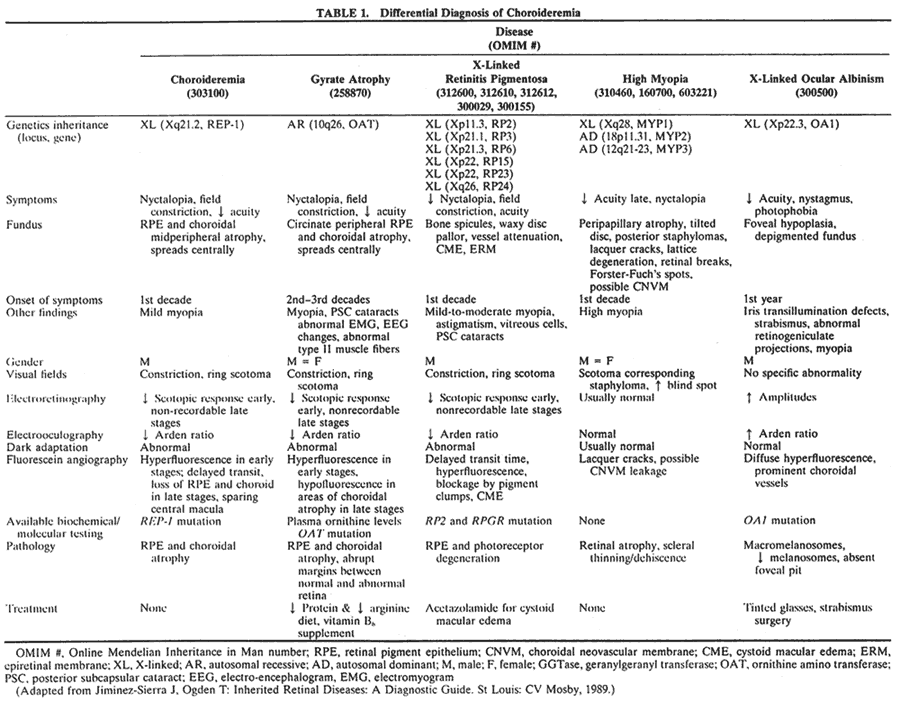
The main differential diagnosis for a diffuse choroidal atrophy includes choroideremia, gyrate atrophy59–61 (Fig. 7A), and advanced X-linked retiniti pigmentosa62–67 (see Fig. 7B). High myopia68–70 (see Fig. 7C) and X-linked ocular albinism71,72 (see Fig. 7D) can sometimes be difficult to distinguish from choroideremia as well. The features of these conditions are shown in Table 1.

Fig. 7. Differential diagnosis of choroideremia. A. Gyrate atrophy. B. X-linked retinitis pigmentosa.C. High myopia. D. X-linked ocular albinism.


Choroideremia has been associated with obesity and deafness,73 hypopituitarism,74 and mental retardation.75–77 These associations are probably due to contiguous gene deletions on the X-chromosome and not directly related to the genetic defect found in choroideremia.

Back to Top
REFERENCES

1. Mauthner H: Ein Fall von Choroideremi. Berl Nat Med Innsbruck 2:191, 1872

2. McCulloch C, McCulloch R: A hereditary and clinical study of choroideremia. Trans Am Acad Ophthalmol Otolaryngol 52:160, 1948

3. Pameyer J, Waardenburg P, Henkes H: Choroideremia. Br J Ophthalmol 44:724, 1960

4. van den Hurk JA, Schwartz M, van Bokhoven H et al: Molecular basis of choroideremia (CHM): Mutations involving the Rab escort protein-1 (REP-1) gene. Hum Mutat 9:110, 1997

5. van den Hurk JA, Hendriks W, van de Pol DJ et al: Mouse choroideremia gene mutation causes photoreceptor cell degeneration and is not transmitted through the female germline. Hum Mol Genet 6:851, 1997

6. McDonald I: Choroideremia. New York: Oxford University Press, 1998

7. Abramson D, Fishman H: Chromosomal studies in choroi-deremia. J Pediatr Ophthalmol Strabismus 10:178, 1973

8. Gordon D: Choroideremia. Med Radiogr Photogr 28:110, 1952

9. Karna J: Choroideremia: A clinical and genetic study of 84 Finnish patients and 126 female carriers. Acta Ophthalmol Suppl 176:1, 1986

10. Heckenlively J: The frequency of posterior subcapsular cataract in the hereditary retinal degenerations. Am J Ophthalmol 93:733, 1982

11. Cherkunov B, Panormova N: Progressive chorioretinal dystrophy (choroideremia) traced down in three generations. Vestn Oftalmol 1:70, 1967

12. Krill A: Krill's Hereditary Retinal and Choroidal Diseases. Hagerstown, MD: Harper & Row, 1977

13. Heckenlively J: Choroideremia. In Heckenlively JR, Arden GB, (eds): Principles and Practice of Clinical Electrophysiology of Vision. St Louis: Mosby—Year Book, 1991

14. Sieving PA, Niffenegger JH, Berson EL: Electroretinographic findings in selected pedigrees with choroideremia. Am J Ophthalmol 101:361, 1986

15. Kurstjens J: Choroideremia and gyrate atrophy of the choroids and retina. Doc Ophthalmol 19:1, 1965

16. Krill A, Archer D: Classification of the choroidal dystrophies. Am J Ophthalmol 72:187, 1971

17. Forsius H, Hyvarinen L, Nieminen H et al: Fluorescein and indocyanine green fluorescence angiography in the study of affected males and in female carriers with choroideremia. Acta Ophthalmol 55:459, 1977

18. Noble KG, Carr RE, Siegel IM: Fluorescein angiography of the hereditary choroidal dystrophies. Br J Ophthalmol 61:43, 1977

19. Gass J: Stereoscopic Atlas of Macular Disease: Diagnosis and Treatment. St Louis: Mosby, 1997

20. Ohba N: Choroideremia: Study of two Japanese families. Acta Soc Ophthalmol Japan 78:116, 1974

21. Cameron J, Fine B, Shapiro I: Histopathologic observations in choroideremia with emphasis on vascular changes of the uveal tract. Ophthalmology 94:187, 1987

22. Rodrigues M, Ballintine E, Wiggert B et al: Choroideremia: A clinical, electron microscopic and biochemical report. Ophthalmology 91:873, 1984

23. Goedbloed J: Mode of inheritance of choroideremia. Ophthalmologica 104:308, 1942

24. Waardenburg P: Choroideremia als Erbuerknal (zur atrophic). Acta Ophthalmol 20:235, 1942

25. Lyon M: Sex chromatin and gene action in the mammalian X-chromosome. Am J Hum Genet 14:135, 1962

26. Lewis RA, Nussbaum RL, Ferrell R: Mapping X-linked ophthalmic diseases: Provisional assignment of the locus for choroideremia to Xq13-q24. Ophthalmology 92:800, 1985

27. Cremers F, van de Pol O, van Kerkhoff L et al: Cloning of a gene that is rearranged in patients with choroideremia. Nature 347:674, 1990

28. Merry DE, Janne PA, Landers JE et al: Isolation of a candidate gene for choroideremia. Proc Natl Acad Sci USA 89:2135, 1992

29. Seabra MC, Brown MS, Goldstein JL: Retinal degeneration in choroideremia: deficiency of rab geranylgeranyl transferase. Science 259:377, 1993

30. Tolmachova T, Ramalho JS, Anant JS et al: Cloning, mapping and characterization of the human RAB27A gene. Gene 239:109, 1999

31. Seabra MC, Ho YK, Anant JS: Deficient geranylgeranylation of Ram/Rab27 in choroideremia. J Biol Chem 270:24420, 1995

32. Cremers F, Molloy C, van de Pol D et al: An autosomal homologue of the choroideremia gene colocalizes with the Usher syndrome type II locus on the distal part of chromosome 1q. Hum Mol Genet 1:71, 1992

33. Cremers FP, Armstrong SA, Seabra MC et al: REP-2, a Rab escort protein encoded by the choroideremia-like gene.J Biol Chem 269:2111, 1994

34. Fraser B, Friedmann A: Choroideremia in a female. BMJ 2:732, 1968

35. Harris G, Miller J: Choroideremia: Visual defects in a heterozygote. Arch Ophthalmol 80:423, 1968

36. Burke M, Choromokos E, Bibler L et al: Choroideremia in a genotypically normal female: A case report. Ophthalmic Pediatr Genet 6:163, 1985

37. Francois J: Importance of electrophysiology in ophthalmogenetics. Ophthalmologica 188:14, 1984

38. Cheng C, Chen M, Hou P: Choroideremia: A study of two families. J Formos Med Assoc 90:1103, 1991

39. Pinckers A, van Aarem A, Brink H: The electrooculogram in heterozygote carriers of Usher syndrome, retinitis pigmentosa, neuronal ceroid lipofuscinosis, senior syndrome and choroideremia. Ophthalmic Genet 15:25, 1994

40. MacDonald I, Chen M, Addison D, Mielke B et al: Histo-pathology of the retinal pigment epithelium of a female carrier of choroideremia. Can J Ophthalmol 32:329, 1997

41. Flannery J, Bird A, Farber D et al: A histopathologic study of a choroideremia carrier. Invest Ophthalmol Vis Sci 31:229, 1990

42. Ghosh M, McCullock C, Parker J: Pathological study ina female carrier of choroideremia. Can J Ophthalmol 23:181, 1988

43. MacDonald IM, Mah DY, Ho YK et al: A practical diagnostic test for choroideremia. Ophthalmology 105:1637, 1998

44. Beaufrere L, Tuffery S, Hamel C et al: The protein truncation test (PTT) as a method of detection for choroideremia mutations. Exp Eye Res 65:849, 1997

45. Hayakawa M, Fujiki K, Hotta Y et al: Visual impairment and REP-1 gene mutations in Japanese choroideremia patients. Ophthalmic Genet 20:107, 1999

46. van Bokhoven H, Schwartz M, Andreasson S et al: Mutation spectrum in the CHM gene of Danish and Swedish choroide-remia patients. Hum Mol Genet 3:1047, 1994

47. Beaufrere L, Tuffery S, Hamel C et al: A novel mutation (S558X) causing choroideremia. Hum Mutat 8:395, 1996

48. Beaufrere L, Tuffery S, Hamel C et al: Rapid genetic diagnosis of females carriers related to patients with choroideremia. (original in French). J Fr Ophtalmol 20:534, 1997

49. Beaufrere L, Tuffery S, Hamel C et al: An exonic polymorphism (381A/G) in the choroideremia gene. Genet Couns 8:223, 1997

50. Beaufrere L, Rieu S, Hache J et al: Altered rep-1 expression due to substitution at position + 3 of the IVS13 splice choroi-deremia (CHM) gene. Curr Eye Res 17:726, 1998

51. Beaufrere L, Rieu S, Hache JC et al: Length variations of the poly(T) tract at the exon 3 splice acceptor site of the choroideremia gene. Genet Couns 9:255, 1998

52. Beaufrere L, Girardet A, Arnaud B et al: Update on a diagnostic test for choroideremia: The protein truncation test (PTT) (original in French). J Fr Ophtalmol 21:345, 1998

53. Beaufrere L, Rieu S, Hache JC et al: Altered rep-1 expression due to substitution at position + 3 of the IVS13 splice-donor site of the choroideremia (CHM) gene. Curr Eye Res 17:726, 1998

54. Beaufrere L, Claustres M, Tuffery S: No missense mutation in choroideremia patients analyzed to date. Ophthalmic Genet 20:89, 1999

55. Forsythe P, Maguire A, Fujita R et al: A carboxy-terminal truncation of 99 amino acids resulting from a novel mutation (Arg555 → stop) in the CHM gene leads to choroideremia. Exp Eye Res 64:487, 1997 (Letter)

56. Fujiki K, Hotta Y, Hayakawa M et al: REP-1 gene mutations in Japanese patients with choroideremia. Graefs Arch Clin Exp Ophthalmol 237:735, 1999

57. Pascal O, Donnelly P, Fouanon C et al: A new (old) deletion in the choroideremia gene. Hum Mol Genet 2:1489, 1993

58. Schwartz M, Rosenberg T, van den Hurk JA et al: Identification of mutations in Danish choroideremia families. Hum Mutat 2:43, 1993

59. Jiminez-Sierra J, Ogden T: Inherited Retinal Diseases: A Diagnostic Guide. St Louis: CV Mosby, 1989

60. Mitchell GA, Looney JE, Brody LC et al: Human ornithine-delta-aminotransferase: cDNA cloning and analysis of the structural gene. J Biol Chem 263:14288, 1988

61. McInnes RR, Arshinoff SA, Bell L et al: Hyperornithinaemia and gyrate atrophy of the retina: Improvement of vision during treatment with a low-arginine diet. Lancet 1:513, 1981

62. Schwahn U, Lenzner S, Dong J et al: Positional cloning of the gene for X-linked retinitis pigmentosa 2. Nat Genet 19:327, 1998

63. Meindl A, Dry K, Herrmann K et al: A gene (RPGR) with homology to the RCC1 guanine nucleotide exchange factor is mutated in X-linked retinitis pigmentosa (RP3). Nat Genet 13:35, 1996

64. Ott J, Bhattacharya S, Chen JD et al: Localizing multiple X chromosome-linked retinitis pigmentosa loci using multilocus homogeneity tests. Proc Natl Acad Sci USA 87:701, 1990

65. Mears AJ, Hiriyanna S, Vervoort R et al: Remapping of the RP15 locus for X-linked cone-rod degeneration to Xp11.4-p21.1, and identification of a de novo insertion in the RPGR exon ORF15. Am J Hum Genet 67:1000, 2000

66. Hardcastle AJ, Thiselton DL, Zito I et al: Evidence for a new locus for X-linked retinitis pigmentosa (RP23). Invest Ophthalmol Vis Sci 41:2080, 2000

67. Gieser L, Fujita R, Goring HH et al: A novel locus (RP24) for X-linked retinitis pigmentosa maps to Xq26-27. Am J Hum Genet 63:1439, 1998

68. Young TL, Ronan SM, Alvear AB et al: A second locus for familial high myopia maps to chromosome 12q. Am J Hum Genet 63:1419, 1998

69. Young TL, Ronan SM, Drahozal LA et al: Evidence that a locus for familial high myopia maps to chromosome 18p. Am J Hum Genet 63:109, 1998

70. Schwartz M, Haim M, Skarsholm D: X-linked myopia: Bornholm eye disease: Linkage to DNA markers on the distal part of Xq. Clin Genet 38:281, 1990

71. Bassi MT, Schiaffino MV, Renieri A et al: Cloning of the gene for ocular albinism type 1 from the distal short arm of the X chromosome. Nat Genet 10:13, 1995

72. Schiaffino MV, d'Addio M, Alloni A et al: Ocular albinism: Evidence for a defect in an intracellular signal transduction system. Nat Genet 23:108, 1999

73. Ayazi S: Choroideremia, obesity, and congenital deafness. Am J Ophthal 92:63, 1981

74. Menon RK, Ball WS, Sperling MA: Choroideremia and hypopituitarism: An association. Am J Med Genet 34:511, 1989

75. May M, Colleaux L, Murgia A et al: Molecular analysis of four males with mental retardation and deletions of Xq21 places the putative MR region in Xq21.1 between DXS233 and CHM. Hum Mol Genet 4:1465, 1995

76. Nussbaum RL, Lesko JG, Lewis RA et al: Isolation of anonymous DNA sequences from within a submicroscopic X chromosomal deletion in a patient with choroideremia, deafness, and mental retardation. Proc Natl Acad Sci USA 84:6521, 1987

77. Rosenberg T, Niebuhr E, Yang HM et al: Choroideremia, congenital deafness and mental retardation in a family with an X chromosomal deletion. Ophthalmic Paediatr Genet 8:139, 1987

Back to Top