Chapter 28A
Cytomegalovirus Retinitis
CAROLINE R. BAUMAL and JAY S. DUKER
Main Menu   Table Of Contents

Search

VIRAL CHARACTERISTICS AND EPIDEMIOLOGY
OCULAR MANIFESTATIONS
DIAGNOSIS
TREATMENT
DIFFERENTIAL DIAGNOSIS
REFERENCES

Before 1982, cytomegalovirus (CMV) retinitis was a disease so extraordinarily rare that few ophthalmologists ever observed a case.1 However, because of the acquired immunodeficiency syndrome (AIDS) pandemic, CMV retinitis has now emerged as a common cause of infectious retinitis worldwide. As a consequence, large amounts of data have been compiled concerning the virus's clinical presentation, pattern of infection, disease course, and complications. CMV produces progressive retinal destruction leading to blindness unless anti-CMV treatment is commenced or the underlying cause of systemic immunocompromise is reversed. Multiple new medications displaying in vitro and in vivo anti-CMV efficacy are now available for this previously untreatable infection.2
Back to Top
VIRAL CHARACTERISTICS AND EPIDEMIOLOGY
Cytomegalovirus is a double-stranded DNA virus belonging to the human herpesvirus family.1,3 Other members include herpes simplex virus, varicella zoster virus (VZV), and Epstein-Barr virus. Although CMV is indistinguishable from the other herpesviruses by electron microscopy, its histopathologic appearance, culture characteristics, and antigenic signature usually enable isolation and differentiation with modern laboratory evaluation of infected material.3 Clinical characteristics of systemic CMV infections were initially described in 1905.4 In the 1920s, CMV infections, which were at the time referred to as salivary gland disease, were recognized to be virally mediated.3,5 Later, CMV disease became known as cytomegalic inclusion disease because of the characteristic cellular inclusions the virus produced on light microscopy. One of the major target organs for CMV is the retina.6 Although the virus was a known human pathogen, CMV infection of the retina was not described until the 1950s.7,8 The virus was proven to be the causative agent in presumed CMV retinitis in 1964.8,9

CMV is transmitted systemically by way of infected blood products, bodily secretions, or across the placenta.1 In developed countries, exposure to CMV usually occurs in childhood or early adulthood. Incidence of CMV seropositivity increases with age. By age 35, nearly all persons living in North America have been exposed to CMV and over 80% will continue to show detectable circulating antibodies to the virus.5,10 Incidence of serologically confirmed CMV infections in newborns is between 0.5% and 2.5%.5,10 Fortunately, only 10% of newborns with laboratory evidence of CMV infection show clinical manifestations of the virus. In immunocompetent persons, CMV infection rarely produces clinical disease. However, CMV is an important cause of morbidity and mortality in immunocompromised individuals. Severe systemic CMV infections occur almost exclusively in persons with abnormal cell-mediated immunity and thus affects two major categories of individuals; the immunosuppressed and newborns. After exposure and systemic infection by CMV, whether it occurs during early development or later in life, the virus typically attains a dormant state within specific cells of the host.11 During periods of severe immunosuppression, CMV virus appears to reactivate and infect the retina through hematogenous spread.12

Infection by the human immunodeficiency virus (HIV) is the most common cause of immunosuppression leading to reactivation of CMV and symptomatic infection. Severe CMV infection has also been associated with congenital immunodeficiency syndromes, pharmacologic immunosuppression, organ transplantation, and malignancy and autoimmune disorders.13 CMV is the major cause of ocular morbidity in HIV infected individuals.11,14 Before the introduction of potent antiretroviral therapy for patients with AIDS, the reported estimates for the prevalence of CMV retinitis varied between 10% and 40%.14–17 CMV retinitis was bilateral at presentation in approximately one third of patients.15 CMV was the AIDS-defining diagnosis in about 5% of patients with HIV infections.15,17,18 Certain subpopulations of patients with AIDS appear to have a lower incidence of CMV retinitis.15,19 Pediatric patients with AIDS have a relatively low incidence of CMV retinitis, probably because they are less likely than adults to have been previously exposed to the virus.20,21

Aside from the retina, symptomatic CMV infection can involve the reticuloendothelial system, liver, kidneys, lungs, gastrointestinal system, and central nervous system in immunosuppressedindividuals. It is believed that the presence of active CMV retinitis almost invariably means that a systemic infection is present. However, the systemic disease may be subclinical. The onset of CMV retinitis appears to depend greatly on the CD4+ T-lymphocyte cell count in adults with AIDS. In children, an age-adjusted CD4 count should be considered.21 The risk for CMV retinitis increases as the CD4 cell count decreases. The mean CD4 cell count at the time of CMV diagnosis is typically less than 50 cells/mm3. It is rare for patients with CD4 counts above 200 cells/mm3 to develop the disease, although it may occur in an individual who previously had a CD4 count less than 50 cells/mm3 and is experiencing an initially favorable response with an elevated CD4 count to highly active antiretroviral therapy (HAART).2

Widespread use of HAART in developed countries has led to improvement in survival of patients with AIDS, reduction of all opportunistic infections including CMV retinitis, increased CD4 lymphocyte cell counts, and decreased HIV viral load.22 Treatment with HAART comprises a combination of medications including protease inhibitors alone or in combination with nucleoside analogues. Epidemiologic data gathered since the introduction of HAART are limited. A retrospective study found a decrease in the incidence of primary and relapsing CMV disease, CMV viral load, and CMV antigenemia in AIDS patients associated with the introduction of HAART therapy.23 However, CMV disease may develop or recur in patients in whom HAART has failed. Immune recovery may be incomplete following HAART and may not fully protect against development of CMV retinitis. Thus, CMV retinitis still appears to be a prevalent opportunistic infection in AIDS and it is important to be aware of its features and treatment of this disorder. The new entity of immune recovery uveitis has been characterized in patients with CMV retinitis who are being treated with HAART.24,25 Moreover, discontinuation of anti-CMV therapy in AIDS patients with improved immune function is now an option for this disorder that previously required life-long anti-CMV treatment.26

Back to Top
OCULAR MANIFESTATIONS
At the onset, CMV retinitis may produce few symptoms, giving scant evidence for the infection's potential to cause severe visual loss.11 The presence of symptoms is often related to the retinal location involved. CMV retinitis affecting the posterior pole tends more often to be symptomatic than peripherally located retinitis. Gradual onset of floaters, scintillating scotoma, decreased peripheral or central vision, or metamorphopsia may be noted. However it is not unusual for active infection to be noted initially on routine screening examination. Immediate, acute loss of central vision early in the disease course rarely occurs, although nearly 75% of patients present with disease that is considered immediately sight-threatening by the ophthalmologist.2 Loss of peripheral vision or a scotoma usually causes affected persons to seek medical attention before central visual loss from direct macular destruction. Pain, external injection, or evidence of more than a mild anterior uveitis is extremely unusual with AIDS-associated CMV retinitis.

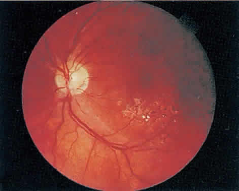
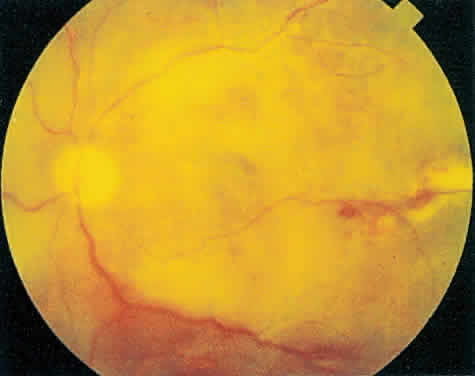
The hallmark lesion of CMV retinitis is a necrotizing, full-thickness retinitis that results in retinal cell destruction. CMV often initially affects retinal tissue adjacent to major retinal blood vessels or the optic disc (Fig. 1). This is consistent with the concept that the virus is spread to the retina hematogenously. In most cases, the pattern of infection is classic and distinctive, making clinical diagnosis straightforward. The area of active retinitis has a granular, dirty-white appearance. As the virus attacks the endothelial cells of blood vessels, hemorrhage is common. Advancement in the retinitis by both direct cell-to-cell transmission as well as spread by way of adjacent satellite lesions can be seen. Except for cases in which retinitis is acute, it is common to see areas of healed retinitis beside areas of active necrosis. Areas of burned-out necrosis show absence of any retinal tissue, whereas the underlying retinal pigment epithelium assumes a “salt and pepper” appearance. CMV retinitis can present initially as either large areas of retinal necrosis with hemorrhage or one or more small, focal areas of retinal whitening.1,12,14 These small, focal lesions may on occasion be confused with cotton-wool spots or lesions of toxoplasmosis.5,27 Unlike cotton-wool spots, focal areas of CMV may appear outside the posterior pole. These early, focal infiltrates of CMV may not be associated with retinal hemorrhages or vitreous cells.

Fig. 1. Typical appearance of peripapillary cytomegalovirus retinitis with hemorrhage and exudate. Despite the proximity to the optic nerve, the visual acuity was 20/20 (6/6) and the patient had no symptoms.

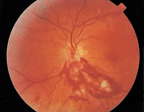
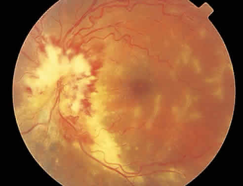
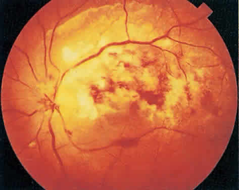
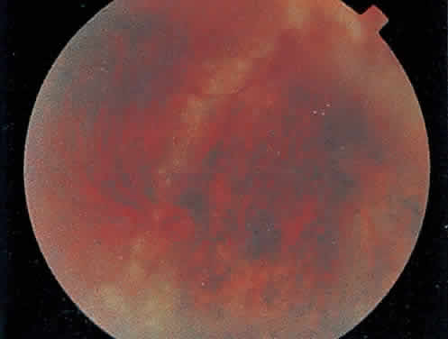
Over a course that usually spans weeks, infiltrates of CMV tend to assume two different patterns of clinical disease.11,12 The first pattern is called hemorrhagic and is characterized by broad geographic zones of retinal whitening. These large, geographic lesions are usually in close proximity to a major retinal blood vessel or the optic nerve. Satellite lesions are common. When the retinal necrosis associated with CMV retinitis becomes widespread, it is almost invariably associated with retinal hemorrhages. Although the border between necrotic and unaffected retina is sharply demarcated, the border itself appears irregular and jagged. Exudation into the retina or subretinal space may be seen, adding to the granular appearance of the retinitis. Juxtaposition of large zones of white, granular necrosis with those of red retinal hemorrhage has led this appearance of CMV retinitis to be described as either “pizza-pie” or “cheese and ketchup.” The retinal blood vessels, both arteries and veins, in the areas of necrosis commonly appear sheathed, secondary to a vasculitis. As a consequence, secondary retinal vascular occlusions, especially branch retinal vein obstructions, may occur in the course of CMV retinitis. Immune-mediated vascular damage may play a role in the vasculitis.12 Central healing of these lesions will occur as the infection progresses. Avasculitis resembling “frosted branch angiitis” hasbeen reported (Fig. 2).28 A second pattern of CMVretinitis has been labeled “granular” or “brushfireborder.” In this appearance, the focal granular infiltrates enlarge slowly across a line, leaving ever-increasing areas of destroyed retina and atrophic retinal pigment epithelium behind. Hemorrhages and vitreous cells are a less prominent feature. There appears to be direct cell-to-cell transfer of infected virions in this pattern of infection (Figs. 3 and 4). The brushfire border is commonly seen in CMV retinitis lesions anterior to the equator (Fig. 5). The significance, if any, of these two clinical patterns of CMV retinitis is not known, and, in some eyes, both patterns of disease can be seen simultaneously or in sequence. Progression of retinitis has been defined in clinical trials as movement of a lesion border at least 750 μm along a front that is 750 μm or more in length, development of a new CMV lesion in a previously involved eye or in the uninvolved fellow eye of a patient with baseline unilateral disease.29 Without treatment or improvement in the host's immune system, CMV retinitis is a relentless, slowly progressive infection resulting in blindness caused by total retinal necrosis, retinal detachment, or optic nerve involvement, in any combination.

Fig. 2. Active cytomegalovirus retinitis concentrated around the optic nerve. Temporal to the fovea, there is perivascular whitening with a frosted branch angiitis appearance.

Fig. 3. Active cytomegalovirus retinitis adjacent to the optic nerve. Visual acuity was counting fingers.

Fig. 4. Three months later the retinitis has completely destroyed the retina leaving a visual acuity of no light perception.

Fig. 5. Brushfire border pattern of cytomegalovirus retinitis inferonasally in a left eye. Temporal to the line of active retinitis there is a “salt and pepper” appearance to the retinal pigment epithelium secondary to the burned-out retinitis. Uninvolved retina is seen nasally.

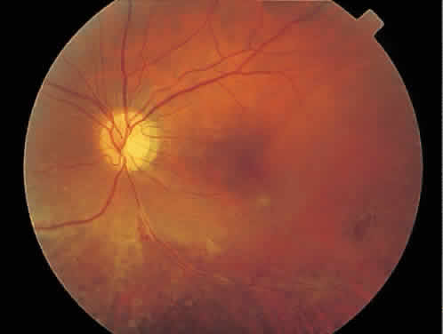
CMV infection can affect the optic nerve either directly or by extension from adjacent retinitis (see Fig. 2).30–32 When direct involvement occurs, optic neuritis with profound, irreversible visual loss usually develops. Several cases of CMV optic neuritis associated with adjacent retinitis have been treated successfully. Exudative retinal detachment can occur during the course of CMV retinitis as well.4,12,30 The subretinal fluid is seen primarily inferiorly in the fundus and shifts with position. No retinal break will be evident; however, in areas of extremely thin, atrophic retina, it can be quite difficult to determine whether a full-thickness defect is present. Exudative retinal detachment associated with CMV is usually nonprogressive and may respond to ganciclovir therapy.

Other features may include a mild to moderate anterior chamber cell and flare reaction. A hypopyon has rarely been reported in a renal transplant patient.4 Although mild vitreous cells are almost universally present, a florid vitritis severe enough to result in media opacity rarely develops from isolated CMV retinitis. In one patient who was immunosuppressed as a result of systemic corticosteroid therapy, a severe panuveitis associated with CMV retinitis was reported.32 More recently, an entity called immune recovery retinitis has been described.26,27 Immune-recovery uveitis is a chronic inflammatory syndrome associated with clinical immune reconstitution in AIDS patients with CMV retinitis who are taking HAART.33–35 It has emerged as an important cause of visual morbidity. Although immune recovery associated with HAART has allowed some patients to discontinue specific anti-CMV therapy, the rejuvenated immune response can be associated with this sight-threatening inflammation in some patients with preexisting CMV retinitis. Ocular features of immune-recovery uveitis include a significant vitritis that is more pronounced than that occurring with primary CMV retinitis. Additional features may include optic disc and macular edema. The mean CD4+ T-lymphocyte count in one study was 393 cells/mm3 at the time of diagnosis.33 Long-term vision-threatening complications related to this persistent inflammation include proliferative vitreoretinopathy, epiretinal membrane formation, posterior subcapsular cataracts, and severe postoperative inflammation.36

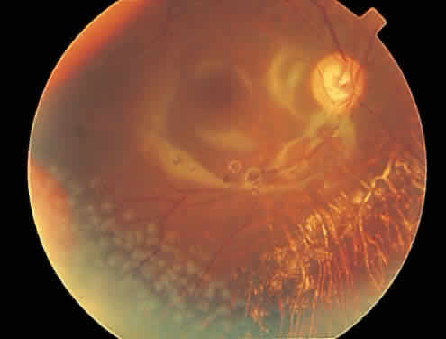
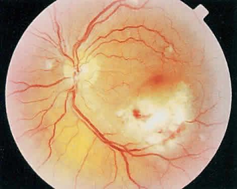
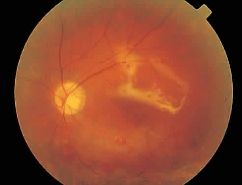
Rhegmatogenous retinal detachment occurs in 10% to 20% of eyes with CMV retinitis.37–39 However, before the AIDS epidemic, there were only five reported cases of this association.10,40 In patients living more than 1 year with CMV retinitis, risk of retinal detachment may be as high as 50%,41 which increases if more than 25% of peripheral retina is involved by disease.41,42 Retinal breaks in eyes with CMV retinitis typically occur within or at the border of necrotic atrophic retina (Fig. 6).43 The ensuing retinal detachments are typically difficult to repair with standard scleral buckling procedures. This is because of the location and number of retinal breaks, the difficulty in visualizing all breaks in necrotic retina, and the high incidence of associated proliferative vitreoretinopathy. In many cases, pars plana vitrectomy and retinal tamponade with silicone oil or long-acting intraocular gas is indicated (Figs. 7 and 8).37,44,45 However, scleral buckling may be considered in small peripheral retinal detachments when the entire involved area can be completely placed on the element. Laser photocoagulation demarcation has also been described to delimit macula-sparing CMV-related retinal detachment.46 Although anatomic success of macular reattachment with surgery is high, the visual results are often limited by the underlying disease process.47 When considering surgical repair of CMV-induced retinal detachment, consideration should be given to the potential for ambulatory vision, the patient's systemic condition and the status of the fellow eye.

Fig. 6. A shallow cytomegalovirus-related retinal detachment is present inferiorly and involves the fovea.

Fig. 7. Repair of the eye in color plate H with silicone oil. Note the silicone oil reflections in the macula.

Fig. 8. Repair of a cytomegalovirus-related rhegmatogenous retinal detachment with pars plana vitrectomy and silicone oil tamponade. Reflections from silicone oil are noted in the macular region. Postoperatively, laser photocoagulation was required inferotemporally to wall off a small area of peripheral subretinal fluid.

Back to Top
DIAGNOSIS
Diagnosis of CMV retinitis is primarily made on a clinical basis.2 In most cases, ophthalmoscopic examination combined with clinical history is sufficient to confirm the diagnosis. Because the disease usually spreads slowly, a period of close careful observation aided by funduscopic photographic documentation may be considered when the diagnosis remain uncertain.11 Although most patients with CMV retinitis have concurrent diffuse systemic CMV infection as well, most of the time they remain systemically asymptomatic. Given that nearly all immunosuppressed patients who are at risk for CMV retinitis will show serologic or culture evidence of the virus in bodily fluids, documented CMV viremia alone, or combined with viruria does not confirm the diagnosis of CMV retinitis.11

Tissue diagnosis of CMV retinitis can be obtained, although an invasive procedure to obtain infected retinal tissue carries potential risk. A retinal biopsy during retinal detachment repair may be performed in the setting of a rhegmatogenous retinal detachment with areas of detached, necrotic retina when the diagnosis is unclear.37,39 Retina biopsy may show cytomegalic cells, although severe tissue necrosis may preclude this finding.

Standard culture of an aqueous or vitreous sample is typically of little assistance in diagnosing CMV retinitis because of the limited involvement of these tissues. Newer techniques such as polymerase chain reaction (PCR) allow detection of CMV DNA from small amounts of intraocular fluid and subsequent diagnosis.48 CMV PCR has demonstrated that aqueous and vitreous specimens can provide accurate markers to differentiate active and inactive CMV retinitis.49 Additionally, these markers were superior to extraocular measures, such as plasma PCR and blood and urine CMV cultures. A direct correlation was identified between the quantity of CMV DNA in aqueous humor or vitreous specimens and the corresponding surface area of active CMV retinitis. This technique may be useful in cases where diagnosis is uncertain or when visualization of the retina is obscured, although it is primarily available as a research tool.

The efficacy of screening asymptomatic patients at risk for CMV retinitis has not been clearly defined. It has been recommended to screen AIDS patients based on their CD4 lymphocyte count. Many patients may be asymptomatic with active retinitis and earlier diagnosis and treatment should result in less extension of CMV into the macula. In addition, reduction of the area of necrotic retina may decrease the incidence of CMV-related retinal detachment.

Back to Top
TREATMENT
Treatment for CMV infections and retinitis has improved significantly over the last decade.50 Principles of therapy include reversal of the underlying cause of immunosuppression if possible and medications with specific anti-CMV activity. In patients who develop CMV retinitis after organ transplantation, reduction of immunosuppressants if medically possible was the treatment before the availability of anti-CMV drugs. In some AIDS patients, this is now possible with the use of HAART. There are scattered reports of antiretroviral therapy (e.g., zidovudine) alone inducing regression of CMV retinitis.51 Before the availability of ganciclovir and fos-carnet, other medical therapies were tried but none proved effective. These included corticosteroids, γ-globulin, antifungal agents, vidarabine, human leukocyte interferon, interferon α, and acyclovir.10,52 In the 1980s, two drugs became available for intravenous treatment of CMV retinitis-ganciclovir and foscarnet. These agents have proven effective initially to halt CMV retinitis. However, these drugs are virostatic, poorly absorbed, and associated with significant systemic toxicities that often limit their use. More recently, four additional treatment options have been approved including oral ganciclovir, intravenous cidofovir, the ganciclovir implant, and intravitreal fomivirsen (Vitravene) injection. These newer remedies avoid the requirement of chronic venous access, daily intravenous therapy and its attendant negative impact on quality of life, and provide the latest option of local therapy.

Ganciclovir is an acyclic nucleoside that is a congener of acyclovir, but it is between 10 and 100 times more effective against CMV.11,15,53 Most isolates of CMV are inhibited by 0.1 to 0.3 μg/ml.53 Virostatic intravenous levels can be achieved with intravenous doses of 2.3 to 5 mg/kg.54 Ganciclovir is also active against the other members of the herpesvirus family (herpes simplex virus, VZV). The recommended intravenous dosage of ganciclovir is 5 to 7.5 mg/kg.2,11 Initially, during the induction phase of treatment, the drug is given twice a day for 2 to 3 weeks. Clearing of active retinitis usually takes several weeks.11 Maintenance therapy usually consists of5 mg/kg once a day, five or seven times per week.2,11,55 If ganciclovir therapy is stopped and the patient remains immunocompromised, recrudescence is usually seen within 4 weeks.11 The initial response rate to intravenous ganciclovir induction varies from 80% to 100%, but reactivation of CMV retinitis during maintenance therapy is common and expected in immunocompromised individuals.2,12,55,56 This has been attributed to low ocular drug bioavailability, progressive decline in immune function and development of CMV resistance to ganciclovir. Resistance to ganciclovir has been associated with a mutation in both the UL97 and UL54 genes.57 Evidence indicates that ganciclovir-treated AIDS patients with CMV retinitis live longer than those who receive no treatment.58 Additionally, patients who show a complete response to ganciclovir appear to live longer than those whose response is only partial.55 Despite ganciclovir treatment, contralateral CMV retinitis may develop in up to 15% of previously unaffected eyes.17 Ganciclovir carries significant bone marrow toxicity. Overall, up to 70% of treated patients may develop some degree of bone marrow suppression.2,55 Severe thrombocytopenia as well as neutropenia can develop, requiring cessation of intravenous therapy. Although bone marrow suppression may limit the dosing, the availability of hematopoietic stimulating factors (i.e., granulocyte-colony stimulating factor, granulocyte monocyte-colony stimulating factor) has permitted improved tolerance of ganciclovir.

The second intravenous agent approved for the treatment of CMV retinitis was foscarnet. Foscarnet (trisodium phosphoformate) is a synthetic, water-soluble pyrophosphate analogue that inhibits in vitro replication of the herpesviruses.2,11,12,39 Its activity is through noncompetitive binding to the exchange site of viral DNA polymerase, thereby rendering it inactive. Foscarnet lacks the bone marrow toxicity of ganciclovir, allowing for its concurrent use with zidovudine. It requires extensive hydration during intravenous therapy, and it may induce nephrotoxicity and seizures.2,59 The induction regimen for foscarnet is 60 mg/kg every 8 hours or 90 mg/kg twice daily.2,59 It is usually initiated for 2 to 3 weeks, followed by maintenance therapy at 90 to 120 mg/kg/day. The efficacy of foscarnet to induce regression of retinitis and maintain CMV inactivity mirrors that of intravenous ganciclovir very closely.2,60 The Studies of the Ocular Complications of AIDS (SOCA) Research Group showed that the two drugs appear equivalent in controlling CMV retinitis and preserving vision, although foscarnet may be associated with longer survival than ganciclovir.60 In general, however, ganciclovir appears to be better tolerated than foscarnet for long-term therapy.

Besides their side effects, both ganciclovir and foscarnet have important disadvantages. Neither is viricidal, so their use must be maintained for as long as the affected individual remains immunosuppressed.2,55 The relapse rate during maintenance therapy is high. Relapses may be controlled by reinduction with either agent or a combination of both intravenous ganciclovir and foscarnet. The improved efficacy of this combination in controlling relapsing retinitis is counteracted by its side effects and negative impact on quality of life measures.61 For this reason, combination therapy is reserved for severely resistant cases. An alternative to combination intravenous therapy that is now available would be placement of a ganciclovir intraocular implant combined with intravenous or intravitreal foscarnet. Either foscarnet or ganciclovir is typically administered intravenously for induction. This often requires placement of a permanent intravenous access system with the subsequent risk of infection. Finally, both drugs are costly and frequent dosing is time consuming and inconvenient for the patient.

An oral form of ganciclovir became available in the mid 1990s. It is used as maintenance therapy in patients who respond well to initial intravenous induction with ganciclovir. However, it has poor ocular bioavailability. This necessitates the use of large doses (ranging between 3 to 6 g per day) and the median time interval to progression of retinitis is less with oral ganciclovir than the intravenous form (29 versus 49 days respectively).62 The risk of developing CMV retinitis in the contralateral eye is greater with oral than with intravenous ganciclovir. Oral ganciclovir appears to play a role in decreasing the risk of fellow eye retinitis in patients with unilateral CMV retinitis who are treated with local therapy such as the ganciclovir implant.63 A prodrug of oral ganciclovir with improved bioavailability is currently under study.

Cidofovir (also known as HPMPC) is an antiviral nucleotide analogue with significant activity against CMV and other herpesviruses. Cidofovir has a long intracellular half-life that allows for a prolonged interval of 2 weeks between intravenous maintenance doses, compared with daily administration of intravenous ganciclovir and foscarnet.64 The efficacy of intravenous cidofovir has been demonstrated in patients with AIDS and previously untreated CMV retinitis in multicenter randomized trials, and in a dose-finding study of cidofovir in patients with AIDS and previously treated relapsing CMV retinitis.65,66 Clinical trials have involved relatively small numbers of patients and no studies have beenconducted directly comparing intravenous cido-fovir with either ganciclovir or foscarnet. Indirectcomparisons of clinical trial data suggest that in-travenous cidofovir may have similar efficacy tointravenous ganciclovir or foscarnet in delaying progression of CMV retinitis. However, such comparisons must be made cautiously because of differences in study design and examination techniques.

Despite this, intravenous cidofovir is less invasive and more convenient for patients because of its prolonged dosage interval. In addition, an indwelling central venous catheter is not required. The long dosage interval for cidofovir may also have favorable implications regarding treatment costs and quality of life. The major treatment-limiting adverse event associated with intravenous cidofovir is potentially irreversible nephrotoxicity. Close monitoring of renal function, hydration, and simultaneous administration of probenecid on treatment days are required. An anterior uveitis syndrome has been reported in up to 40% of patients 67 and a small number of patients have developed hypotony.

Cidofovir and ganciclovir appear to have a synergistic effect inhibiting CMV replication in vitro. A phase I study suggested that intravenous cidofovir (5 mg/kg intravenously every 2 weeks) combined with oral ganciclovir might enhance treatment efficacy in AIDS-related CMV retinitis.68 Notably, this regimen does not require indwelling venous catheter access. Because all the intravenous treatment options for CMV retinitis are associated with potentially serious adverse events, selection of pharmacotherapy should be individualized and depends several factors including lesion characteristics, patient quality of life issues, and efficacy and tolerability profiles of available therapies. While receiving maintenance intravenous therapy, patients are typically examined every 4 weeks for evidence of reactivation of the CMV retinitis. When this occurs, options for further treatment include reinduction, medication switch, combination therapy, or adjunctive local therapy. Fundus photographs are invaluable for assessing the activity of the retinitis from visit to visit and should be repeated whenever a change is suspected.

Local intraocular treatments as alternatives to systemic medications have been a significant advance in management of CMV retinitis. Local ocular therapy has proven effective and provides significant advantages over systemic therapy with regards to control of retinitis, prevention of progression, and avoidance of bone marrow and renal toxicity. Disadvantages of local therapy include the risk of CMV retinitis in the contralateral eye and the lack of protection against systemic extraocular CMV infection.

Animal experiments demonstrated that intravitreal injections in excess of 400 μg of ganciclovir and 1200 μg of foscarnet are nontoxic to the retina.69,70 Intravitreal injection of either ganciclovir or foscarnet, although an unapproved modality, has been used successfully in some patients, especially those with recurrent or refractory disease. Long-term control of CMV retinitis control through repeated intravitreal injections has been evaluated.54,71,72 The published dose of intravitreal ganciclovir is 200 to 400 μg, although many clinicians now use a dose of 2000 μg in 0.1-ml sterile normal saline. This appears to control active CMV retinitis in about 80% of patients. During the induction phase, injections are given three times per week, followed by injections given once or twice per week during long-term maintenance therapy. Multiple injections are required because of the short intraocular drug half-life. About 30% of treated patients develop recurrent disease on this regimen.71,72 As an alternative in patients who are resistant to ganciclovir, intravitreal foscarnet at a dose of up to 2400 μg in 0.1 ml of sterile normal saline may control CMV retinitis.73 No retinal toxicity has been associated with the doses mentioned here. Potentially vision-threatening complications associated with this method such as exogenous endophthalmitis, vitreous hemorrhage, and retinal detachment are uncommon. However, chronic intravitreal injections are not well tolerated because of the inconvenience of frequent injections. Cidofovir has also been administered by intravitreal injection and it has the advantage of a longer intracellular half-life, which would allow less frequent injections. However, it has been associated with multiple side effects including transient or chronic hypotony with permanent visual loss and uveitis. Further study is needed to determine the safety of this agent for intraocular injection and the effective intraocular dose.74

An intraocular ganciclovir implant that delivers this drug in a sustained release fashion is well tolerated and very effective against CMV retinitis. Advantages include less frequent injection than the above intravitreal agents and a constant rate of drug release into the vitreous. This intraocular implant was approved by the U.S. Food and Drug Administration (FDA) in 1996 (Fig. 9).75,76 The implant consists of a pelletized form of ganciclovir covered by a semipermeable membrane. It contains approximately 5 mg of drug, with a release rate of 1.5 μg/hr and the drug lasts between 6 to 8 months. It is placed surgically into the vitreous cavity through a pars plana incision and releases ganciclovir linearly in a time-released fashion (Fig. 10).77,78 The time to progression of CMV retinitis is significantly longer with the ganciclovir implant than with intravenous therapy, but there is also a higher rate of contralateral eye retinitis and systemic CMV disease with the implant (Figs. 11 and 12).79 The implant has been used to treat primary, recurrent, and bilateral retinitis with implantation in each eye. It has also been effective in eyes with silicone oil tamponade for CMV-related retinal detachment.80–82 Complications are uncommon but include macular edema, vitreous hemorrhage, and retina detachment, especially if CMV infection involved more than 25% of the retina. The device can be replaced at approximately 6 months when it is depleted of drug, but some clinicians prefer to observe for early signs of recurrent CMV activity before replacement.83 In addition, multiple implants can be placed in one eye rather than exchanging the implant when it is depleted of drug. A very rare complication was two cases of separation of the ganciclovir medication pellet from the tab of the implant when it was removed.84

Fig. 9. Close-up of the intravitreal sustained-release ganciclovir implant.

Fig. 10. Intravitreal sustained-release ganciclovir implant in situ. (Courtesy of Michael Robinson.)

Fig. 11. Active cytomegalovirus retinitis just inferior to the macula. The visual acuity is 20/30 (6/9).

Fig. 12. Three months after implantation of an intravitreal sustained-release ganciclovir implant, the retinitis is completely healed and the vision is 20/25 (6/7.5).

The beneficial effect of HAART on the immunologic status and survival in some patients with HIV has altered the incidence of primary and relapsing CMV retinitis. Thus, clinical management of CMV retinitis and the role of the ganciclovir implant are changing.85 The International AIDS Society (U.S.A. panel) concluded that optimal use of the ganciclovir implant and discontinuation of systemic therapy in selected patients with improved immunity due to HAART may result in better long-term visual outcomes.86 In patients who remain chronically immunocompromised and are treated with local therapy, systemic anti-CMV therapy in the form of oral ganciclovir is recommended to avoid CMV infection in the fellow eye as well as symptomatic extraocular CMV disease.63

Fomivirsen (also known as Vitravene or ISIS 2922, manufactured by Ciba Vision) is an antisense oligonucleotide, which specifically inhibits replication of human CMV. It achieves this by binding to complementary sequences on messenger RNA transcribed from a major transcriptional unit of the virus.87,88 It is administered as an intravitreal injection and is an alternative treatment for refractory CMV retinitis. It has a longer intravitreal elimination half-life than either ganciclovir or foscarnet, allowing injections at intervals of 1 week for induction and every 2 weeks for maintenance. Commonly reported side effects include increased intraocular pressure and mild to moderate intraocular inflammation, which is generally transient and reversible with topical steroid drops. Two cases of bull's-eye maculopathy have been reported that were reversible with discontinuation of therapy; this did not appear to affect vision.89

Development of new anti-CMV agents has been slowed by the decreased incidence of the disease. Valganciclovir, a prodrug of ganciclovir, offers excellent oral bioavailability and is the closest to approval of the new anti-CMV drugs. High ganciclovir blood levels are achieved without the complications associated with chronic intravenous access.

Factors to consider in selecting the mode of therapy include the patient's potential for immunologic improvement, the location and severity of CMV retinitis, and the risks, costs, and convenience associated with various therapies. Before introduction of HAART, lifelong anti-CMV therapy was required to prevent progression of CMV retinal disease and subsequent loss of vision in AIDS. The beneficial effect of HAART on the immunologic status of some patients has made the role of maintenance anti-CMV treatment less clear. Maintenance anti-CMV medications have been safely stopped in some small series of AIDS patients with stable inactive CMV retinitis and elevated CD4+ cell counts (above 100 cells/μl) without reactivation of CMV retinitis.26,90,91 Thus, immune recovery following potent HAART may be effective to control a major opportunistic infection, even in patients with a history of previous severe immunosuppression. Patients may be at risk for reactivation of CMV retinitis despite initial favorable response to HAART if their CD4 lymphocyte count falls below 50 cells/mm3.92

Back to Top
DIFFERENTIAL DIAGNOSIS
Any entity that produces a focal or diffuse retinitis with or without retinal hemorrhage in an immunosuppressed host should be considered in the differential diagnosis of CMV retinitis. Because immunosuppressed patients can also develop a myriad of other opportunistic ocular infections, the treatments for which may differ dramatically from CMV retinitis, differentiation is crucial.11,12 For the most part, the clinical history and a careful ophthalmoscopic examination of both eyes will be sufficient to identify CMV as the offending pathogen. In unusual cases, a period of close observation, an empiric treatment trial with anti-CMV therapy, or even a vitreous or retinal biopsy may be indicated.

HIV retinopathy is the entity most likely to be confused with early CMV retinitis.27 It is the most common ocular manifestation of patients with AIDS, occurring in about two thirds of those diagnosed with the disease.15,18 HIV retinopathy consists of multiple, cotton-wool spots, scattered retinal hemorrhages, and is usually bilateral. If these areas of retinal whitening resemble CMV, close observation over a period of days to weeks with documentation by photography is recommended. CMV retinitis will invariably progress, whereas cotton wool spots eventually resolve. HIV retinopathy rarely produces visual loss.

The most important posterior segment infections that may be confused with CMV retinitis are toxoplasmosis, syphilis, and infection with the other herpesviruses (herpes simplex virus, VZV). Toxoplasmosis is often a nonopportunistic infection and generally presents as a focal retinitis with more vitreous cells and less retinal hemorrhages than CMV retinitis does. However, in immunocompromised hosts, Toxoplasma infection can produce a severe, diffuse retinitis.93 Serologic diagnosis may be helpful with toxoplasmosis. Nearly one third of HIV patients have CNS toxoplasmosis concurrently. Ocular syphilis can also mimic CMV retinitis, although the lesions of secondary syphilis are usually a choroiditis rather than a retinitis.94 Serologic testing is invaluable in diagnosing syphilis. Opportunistic infections resulting in the clinical syndrome of multifocal choroiditis such as Pneumocystis carinii infection, histoplasmosis, cryptococcosis, and atypical Mycobacteria should rarely, if ever, be confused with CMV retinitis on ophthalmoscopic examination.

The acute retinal necrosis (ARN) syndrome due to VZV or, more rarely, herpes simplex virus occurs in both healthy persons and the immunosuppressed, including those with AIDS.95 ARN differs from CMV retinitis in that it is typically peripheral in location and much more rapid in course. In addition, vitreous cells are a prominent feature of ARN. A specific type of rapidly progressive ARN with little vitreous reaction associated with VZV in AIDS patients has been described.96 In cases of suspected ARN, intravenous acyclovir is the recommended treatment. If no response is seen, it is reasonable to try foscarnet because it is the drug of choice for acyclovir-resistant herpesviruses. Acyclovir is rarely effective against CMV.

Back to Top
REFERENCES

1. Hennis HL, Scott AA, Apple DJ: Cytomegalovirus retinitis. Surv Ophthalmol 34:193, 1989

2. Studies of the Ocular Complications of AIDS Research Group: Mortality in patients with the acquired immune deficiency syndrome treated with either foscarnet or ganciclovir for cytomegalovirus retinitis. N Engl J Med 326:213, 1992

3. Freidman AH, Orellana J, Freeman WR et al: Cytomegalovirus retinitis: A manifestation of the acquired immune deficiency syndrome (AIDS). Br J Ophthalmol 67:372, 1983

4. Chumbley LC, Robertson DM, Smith TF et al: Adult cytomegalovirus inclusion retino-uveitis. Am J Ophthalmol 80:807, 1975

5. Nicholson DH: Cytomegalovirus infections of the retina. Int Ophthalmol Clinic 15:151, 1975

6. Murray HW, Knox DL, Green WR et al: Cytomegalovirus retinitis in adults: A manifestation of disseminated viral infection. Am J Med 63:574, 1977

7. Christensen L, Beeman HW, Allen A: Cytomegalic inclusion disease. Arch Ophthalmol 57:90, 1957

8. Foester HW: Uveitis symposium. Surv Ophthalmol 4:296, 1959

9. Smith ME: Retinal involvement in adult cytomegalic inclusion disease. Arch Ophthalmol 72:44, 1964

10. Meredith TA, Aaberg TM, Reeser FH: Rhegmatogenous retinal detachment complicating cytomegalovirus retinitis. Am J Ophthalmol 87:793, 1979

11. Bloom JN, Palestine AG: The diagnosis of cytomegalovirus retinitis. Ann Intern Med 109:963, 1988

12. Palestine AG: Clinical aspects of cytomegalovirus retinitis. Rev Infect Dis 10:515, 1988

13. Baumal CR, Levin AV, Kavalec C et al: Screening for cytomegalovirus retinitis in children. Arch Pediatr Adolesc Med 150:1186, 1996

14. Schuman JS, Orellana J, Friedman AH et al: Acquired immunodeficiency syndrome (AIDS). Surv Ophthalmol 31:384, 1987

15. Jabs DA, Green WR, Fox R et al: Ocular manifestations of acquired immune deficiency syndrome. Ophthalmology 96:1092, 1989

16. Freeman SR, Lerner CW, Mines JA et al: A prospective study of the ophthalmologic findings in the acquired immune deficiency syndrome. Am J Ophthalmol 97:133, 1984

17. Gross JS, Bozzette SA, Mathews WC et al: Longitudinal study of cytomegalovirus retinitis in acquired immune deficiency syndrome. Ophthalmology 97:681, 1990

18. Sison RF, Holland GN, MacArthur LJ et al: Cytomegalovirus retinopathy as the initial manifestation of the acquired immunodeficiency syndrome. Am J Ophthalmol 112:243, 1991

19. Kestelyn P, Van de Perre P, Rouvoy D et al: A prospective study of the ophthalmologic findings in the acquired immune deficiency in Africa. Am J Ophthalmol 100:230, 1985

20. Levin AV, Zeichner S, Duker JS et al: Cytomegalovirus retinitis in an infant with acquired immune deficiency syndrome (AIDS). Pediatrics 84:683, 1989

21. Baumal CR, Levin AV, Read S: Cytomegalovirus retinitis in immunosuppressed children. Am J Ophthalmol 1999;106:301

22. Deayton JR, Wilson P, Sabin CA et al: Changes in the natural history of cytomegalovirus retinitis following the introduction of highly active antiretroviral therapy. AIDS 14:1163, 2000

23. Varani S, Spezzacatena P, Manfredi R et al: The incidence of cytomegalovirus (CMV) antigenemia and CMV disease is reduced by highly active antiretroviral therapy. Eur J Epidemiol 16:433, 2000

24. Karavellas MP, Lowder CY, MacDonald JC et al: Immune recovery vitritis associated with inactive cytomegalovirus retinitis. Arch Ophthalmol 116:169, 1998

25. Zegans ME, Walton RC, Holland GN et al: Transient vitreous inflammatory reactions associated with combination antiretroviral therapy in patients with AIDS and cytomegalovirus retinitis. Am J Ophthalmol 125:292, 1998

26. Vrabec TR, Baldassano VF, Whitcup SM: Discontinuation of maintenance therapy in patients with quiescent cytomegalovirus retinitis and elevated CD4+ counts. Ophthalmology 105:1259, 1998

27. O'Donnell JJ, Jacobson MA: Cotton-wool spots and cytomegalovirus retinitis in AIDS. Int Ophthalmol Clin 29:105, 1989

28. Mansour AM, Li HK: Frosted retinal periphlebitis in the acquired immunodeficiency syndrome. Ophthalmologica 207:182, 1993

29. Holbrook JT, Davis MD, Hubbard LD et al: Risk factors for advancement of cytomegalovirus retinitis in patients with acquire immunodeficiency syndrome: Studies of the Ocular Complications of AIDS Research Group. Arch Ophthalmol 11:1196, 2000

30. Gross JG, Sadun AA, Wiley CA et al: Severe visual loss related to isolated peripapillary retinal and optic nerve cytomegalovirus infection. Am J Ophthalmol 108:691, 1989

31. Grossniklaus HE, Frank KE, Tomsak RL: Cytomegalovirus retinitis and optic neuritis in acquired immune deficiency syndrome. Ophthalmology 94:1601, 1987

32. Berger BB, Weinberg RS, Tessler HH et al: Bilateral cytomegalovirus panuveitis after high-dose corticosteroid therapy. Am J Ophthalmol 88:1020, 1979

33. Robinson MR, Reed G, Csaky KG et al: Immune-recovery uveitis in patients with cytomegalovirus retinitis taking highly active antiretroviral therapy. Am J Ophthalmol 130:49, 2000

34. Kuppermann BD, Holland GN: Immune recovery uveitis. Am J Ophthalmol 130:103, 2000

35. MacDonald JC, Karavellas MP, Torriani FJ et al: Highly active antiretroviral therapy-related immune recovery in AIDS patients with cytomegalovirus retinitis. Ophthalmology 107:877, 2000

36. Karavellas MP, Song M, Macdonald JC et al: Long-term posterior and anterior segment complications of immune recovery uveitis associated with cytomegalovirus retinitis. Am J Ophthalmol 130:57, 2000

37. Freeman WR, Henderly DE, Wan WL et al: Prevalence, pathophysiology, and treatment of rhegmatogenous retinal detachment in treated cytomegalovirus retinitis. Am J Ophthalmol 103:527, 1987

38. Freeman WR, Schneiderman TE: Invasive posterior segment diagnostic procedures in immunosuppressed patients. Int Ophthalmol Clinic 29:119, 1989

39. Holland GN, Sidikaro Y, Kreiger AE et al: Treatment of cytomegalovirus retinopathy with ganciclovir. Ophthalmology 94:815, 1987

40. Broughton WL, Cupples HP, Parver LM: Bilateral retinal detachment following cytomegalovirus retinitis. Arch Ophthalmol 96:618, 1978

41. Studies of Ocular Complications of AIDS (SOCA) Research Group in Collaboration with the AIDS Clinical Trials Group (ACTG): Rhegmatogenous retinal detachment in patients with cytomegalovirus retinitis: The Foscarnet-Ganciclovir Cytomegalovirus Retinitis Trial. Am J Ophthalmol 124:61, 1997

42. Freeman WR, Friedberg DN, Berry C et al: Risk factors for development of rhegmatogenous retinal detachment in patients with cytomegalovirus retinitis. Am J Ophthalmol 116:713, 1993

43. Baumal CR, Reichel E: Management of cytomegalovirus-related rhegmatogenous retinal detachment. Ophthalmic Surg Lasers 29:916, 1998

44. Regillo CD, Vander JF, Duker JS et al: Repair of retinitis-related retinal detachments with silicone oil in patients with acquired immune deficiency syndrome. Am J Ophthalmol 113:21, 1992

45. Dugel PU, Liggett PE, Lee MB et al: Repair of retinal detachment caused by cytomegalovirus retinitis in patients with the acquired immune deficiency syndrome. Am J Ophthalmol 112:235, 1991

46. Vrabec TR: Laser photocoagulation repair of macula-sparing cytomegalovirus-related retinal detachment. Ophthalmology 104:2062, 1997

47. Azen SP, Scott IU, Flynn HW Jr et al: Silicone oil in the repair of complex retinal detachments: A prospective observational multicenter study. Ophthalmology 105:1587, 1998

48. Dodt KK, Jacobsen PH, Hoffman B et al: Development of cytomegalovirus (CMV) disease may be predicted in HIV-infected patients by CMV polymerase chain reaction and the antigenemia test. AIDS 11:F21, 1997

49. Smith IL, Macdonald JC, Freeman WR et al: Cytomegalovirus (CMV) retinitis activity is accurately reflected by the presence and level of CMV DNA in aqueous humor and vitreous. J Infect Dis 179:1249, 1999

50. Hoffman VF, Skiest DJ: Therapeutic developments in cytomegalovirus retinitis. Exp Opin Invest Drugs 9:207, 2000

51. Guyer DR, Jabs DA, Brant AM et al: Regression of cytomegalovirus retinitis with zidovudine. Arch Ophthalmol 107:868, 1989

52. Pollard RB, Egbert PR, Gallaher JG et al: Cytomegalovirus retinitis in immunosuppressed hosts. Ann Intern Med 93:655, 1980

53. Buhles WC, Mastre BJ, Tinker AJ et al: Ganciclovir treatment of life- or sight-threatening cytomegalovirus infection: Experience with 314 immunocompromised patients. Rev Infect Dis 10:495, 1980

54. Ussery FM, Gibson SR, Conklin RH et al: Intravitreal ganciclovir in the treatment of AIDS-associated cytomegalovirus retinitis. Ophthalmology 95:640, 1988

55. Jabs DA, Newman C, De Bustros S et al: Treatment of cytomegalovirus retinitis with ganciclovir. Ophthalmology 94:824, 1987

56. Holland GN, Buhles WC, Mastre B et al: A controlled retrospective study of ganciclovir treatment for cytomegalovirus retinopathy. Arch Ophthalmol 107:1759, 1989

57. Jabs DA, Martin BK, Forman MS et al: Mutations conferring ganciclovir resistance in a cohort of patients with acquired immunodeficiency syndrome and cytomegalovirus retinitis.J Infect Dis 183:333, 2001

58. Holland GN, Sison RF, Jatulis DE et al: Survival of patients with the acquired immune deficiency syndrome after development of cytomegalovirus retinopathy. Ophthalmology 97:204, 1990

59. Lehoang P, Girard B, Robinet M et al: Foscarnet in the treatment of cytomegalovirus retinitis in acquired immune deficiency syndrome. Ophthalmology 96:865, 1989

60. Studies of the Ocular Complications of AIDS Research Group, in collaboration with the AIDS Clinical Trials group: Foscarnet-ganciclovir cytomegalovirus retinitis trial. 4. Visual outcomes. Ophthalmology 101:1250, 1994

61. Studies of Ocular Complications of AIDS Research Group in Collaboration with the AIDS Clinical Trials Group: Combination foscarnet and ganciclovir therapy vs monotherapy for the treatment of relapsed cytomegalovirus retinitis in patients with AIDS: The Cytomegalovirus Retreatment Trial. Arch Ophthalmol 114:23, 1996

62. Drew WL, Ives D, Lalezari JP et al: Oral ganciclovir as maintenance treatment for cytomegalovirus retinitis in patients with AIDS. N Engl J Med 333:615, 1995

63. Martin DF, Kuppermann BD, Wolitz RA et al for the Roche Ganciclovir Study Group: Oral ganciclovir for patients with cytomegalovirus retinitis treated with a ganciclovir implant. N Engl J Med 340:1063, 1999

64. Plosker GL, Noble S. Cidofovir: A review of its use in cytomegalovirus retinitis in patients with AIDS. Drugs 58:325, 1999

65. The Studies of Ocular Complications of AIDS Research Group in collaboration with the AIDS Clinical Trials Group: Long-term follow-up of patients with AIDS treated with parenteral cidofovir for cytomegalovirus retinitis: The HPMPC Peripheral Cytomegalovirus Retinitis Trial. AIDS 14:1571, 2000

66. Studies of Ocular Complications of AIDS Research Group in Collaboration with the AIDS Clinical Trials Group: Parenteral cidofovir for cytomegalovirus retinitis in patients with AIDS: The HPMPC peripheral cytomegalovirus retinitis trial. A randomized, controlled trial. Ann Intern Med 126;264, 1997

67. Davis JL, Taskintuna I, Freeman WR et al: Iritis and hypotony after treatment with intravenous cidofovir for cytomegalovirus retinitis. Arch Ophthalmol 115:733, 1997

68. Jacobson MA, Wilson S, Stanley H et al: Phase I study of combination therapy with intravenous cidofovir and oral ganciclovir for cytomegalovirus retinitis in patients with AIDS. Clin Infect Dis 28:528, 1999

69. Pulido J, Palacio M, Peyman GA et al: Toxicity of intravitreal antiviral drugs. Ophthalmic Surg 15:666, 1984

70. She SC, Peyman GA, Schulman JA: Toxicity of intravitreal injection of foscarnet in the rabbit eye. Int Ophthalmol 12:151, 1988

71. Heinemann MH: Long-term intravitreal ganciclovir therapy for cytomegalovirus retinopathy. Arch Ophthalmol 107:1767, 1989

72. Cantrill HL, Henry K, Melroe NH et al: Treatment of cytomegalovirus retinitis with intravitreal ganciclovir. Ophthalmology 96:367, 1989

73. Diaz-Llopis M, Chipont E, Sanchez S et al: Intravitreal foscarnet for cytomegalovirus retinitis in a patient with acquired immune deficiency syndrome. Am J Ophthalmol 114:742, 1992

74. Taskintuna I, Rahhal FM, Arevalo JF et al: Low-dose intravitreal cidofovir (HPMPC) therapy of cytomegalovirus retinitis in patients with acquired immune deficiency syndrome. Ophthalmology 104:1049, 1997

75. Sanborn GE, Anand R, Torti RE et al: Sustained-release ganciclovir therapy for treatment of cytomegalovirus retinitis. Arch Ophthalmol 110:188, 1992

76. Anand R, Nightingale SD, Fish RH et al: Control of cytomegalovirus retinitis using sustained release of intraocular ganciclovir. Arch Ophthalmol 111:223, 1993

77. Duker JS, Robinson M, Anand R et al: Initial experience with an eight month sustained-release intravitreal ganciclovir implant for the treatment of CMV retinitis associated with AIDS. Ophthalmic Surg Lasers 26:442, 1995

78. Duker JS, Ashton P, Davis JL et al: Long-term successful maintenance of bilateral cytomegalovirus retinitis using exclusively local therapy. Arch Ophthalmol 114:881, 1996

79. Musch DC, Martin DF, Gordon JF, et al: Treatment of CMV retinitis with a sustained-release ganciclovir implant: The Ganciclovir Implant Study Group. N Engl J Med 115:733, 1997

80. Hatton MP, Duker JS, Reichel E et al: Treatment of relapsed CMV retinitis with the sustained-release ganciclovir implant. Retina 18:50, 1998

81. Roth DB, Feuer WJ, Blenke AJ et al: Treatment of recurrent cytomegalovirus retinitis with the ganciclovir implant. Am J Ophthalmol 127:276, 1999

82. McGuire DE, McAulife P, Heinemann MH et al: Efficacy of the ganciclovir implant in the setting of silicone oil vitreous substitute. Retina 20:520, 2000

83. Morley MG, Duker JS, Ashton P et al: Replacing ganciclovir implants. Ophthalmology 102:388, 1995

84. Boyer DS, Posalski J: Potential complication associated with removal of ganciclovir implants. Am J Ophthalmol 127:349, 1999

85. Davis JL, Tabandeh H, Feuer WJ et al: Effect of potent antiretroviral therapy on recurrent cytomegalovirus retinitis treated with the ganciclovir implant. Am J Ophthalmol 127:283, 1999

86. Martin DF, Dunn JP, Davis JL et al: Use of the ganciclovir implant for the treatment of cytomegalovirus retinitis in the era of potent antiretroviral therapy: Recommendations of the International AIDS Society-U.S. A. panel. Am J Ophthalmol 127:329, 1999

87. Amin HI, Ai E, McDonald HR et al: Retinal toxic effects associated with intravitreal forivirsen. Arch Ophthalmol 118:426, 2000

88. Perry CM, Balfour J: Fomivirsen. Drugs 57:375, 1999

89. Stone TW, Jaffe GJ: Reversible bull's-eye maculopathy associated with intravitreal fomivirsen therapy for cytomegalovirus retinitis. Am J Ophthalmol 130:242, 2000

90. Jabs DA, Bolton SG, Dunn JP et al: Discontinuing anticytomegalovirus therapy in patients with immune reconstitution after combination antiretroviral therapy. Am J Ophthalmol 126:817, 1998

91. Whitcup SM, Fortin E, Lindblad AS et al: Discontinuation of anticutomegalovirus therapy in patients with HIV infection and cytomegalovirus retinitis. JAMA 282:1633, 1999

92. Song MK, Karavellas MP, MacDonald JC et al: Characterization of reactivation of cytomegalovirus retinitis in patients healed after treatment with highly active antiretroviral therapy. Retina 20:151, 2000

93. Cochereau-Massin I, LeHoang P, Lautier-Frau M et al: Ocular toxoplasmosis in human immunodeficiency virus-infected patients. Am J Ophthalmol 114:130, 1992

94. McLeish WM, Pulido J, Holland S et al: The ocular manifestations of syphilis in the human immunodeficiency virus type 1-infected host. Ophthalmology 97:196, 1990

95. Duker JS, Blumenkranz MS: Diagnosis and management of the acute retinal necrosis (ARN) syndrome. Surv Ophthalmol 35:327, 1991

96. Forster DJ, Dugel PU, Frangieh GT et al: Rapidly progressive outer retinal necrosis in the acquired immune deficiency syndrome. Arch Ophthalmol 110:341, 1990

Back to Top