Chapter 31 Blunt Trauma ANDREW LAM, WILLIAM E. BENSON and JOHN B. JEFFERS Table Of Contents |
EPIDEMIOLOGY THE NECESSITY OF EYE PROTECTION EVALUATION MECHANISMS OF OCULAR DAMAGE ANTERIOR SEGMENT INJURIES POSTERIOR SEGMENT INJURIES REFERENCES |
This chapter concentrates on posterior segment injuries caused by blunt trauma. Anterior segment injuries, surgical management of penetrating injuries, sports injuries, and injuries in the workplace are covered elsewhere in the textbook. |
EPIDEMIOLOGY |
There are approximately 2.5 million new eye injuries in the United States each year.1 Males are more than four times more likely than females to have ocular injuries and young individuals are more likely than older ones. Blunt objects account for the largest percentage of eye injuries (30%), followed by sharp objects (18%), vehicle crashes (9%), gunshots, nails, BB guns (6% each), fireworks and falls (4% each).1–3 The most common objects to strike the eye are rocks, fists, baseballs, lumber, and fishing weights. In recent years, there has been greater awareness of injuries secondary to bungee cords, paintballs, and airbags.4–9 There are many other rare causes.10–12 Assault and motor vehicle injuries are usually the most severe and are the most likely to result in enucleation.13 The lifetime prevalence of ocular injuries is similar in black and white men, but black men are more likely to sustain a blinding injury.14 |
THE NECESSITY OF EYE PROTECTION |
It is always sad when anyone loses vision from any cause. In the case of sports and occupational injuries, it is doubly sad, because many of them could have been prevented if appropriate, properly fitted, task-specific eye protection had been worn.15,16 Sports injuries account for approximately 100,000 eye injuries per year and approximately 10% to 40% of all eye injuries.17 They are so common because millions of Americans play sports but few wear the appropriate eye protection. The number of serious boxing injuries could probably be reduced if boxers wore proper head gear and thumb-tied gloves.18,19 Similarly, participants in other popular sports, such as squash and racquetball should wear appropriate eye protection.20,21 In Canada, a strict requirement for eye protection has almost totally eliminated blinding injuries in amateur ice hockey, racquetball, and squash players who wear appropriate equipment.22 |
EVALUATION |
In every case of blunt trauma to the eye and orbit it is essential to perform
a complete ophthalmic examination. One of the oddities of blunt
trauma is that an injury that causes minimal signs of damage to one part
of the eye can cause a significant injury in another part of the eye. For
example, a blow to the eye can cause no iritis or hyphema while
causing a giant tear, choroidal rupture, or blowout fracture. Only by
ruling out all possible injuries can the examiner be confident that
none are missed. The examiner should strongly consider an examination
under anesthesia in uncooperative children suspected of being severely
injured. Immediate measurement of visual acuity is important for future decision making and for medico-legal and prognostic purposes. If a Snellen chart is not available, the ability to count fingers or to read a newspaper gives some indication of visual function. The examiner should document the size of the print and the distance at which it is read. A careful assessment of the pupils can be very helpful, because an afferent pupillary defect (APD) is an important predictor of visual outcome.23 In many cases sphincter tears or iritis make this difficult, but the examiner can detect severe retinal or optic nerve damage by using the swinging flashlight test to demonstrate an APD. The pupil in the fellow eye constricts when the light is moved to it from the traumatized one. It is especially important to rule out retinal tears, commotio retinae, choroidal ruptures, and optic nerve involvement. If hyphema is present, however, the fundus examination must be done very gently to avoid precipitating a secondary hemorrhage. Scleral depression and gonioscopy should be avoided for at least 2 to 3 weeks. If a complete fundus examination is difficult because of opaque media, ultrasonography should be repeated every 2 to 3 weeks until it is certain that there is no retinal detachment. All patients must be followed until the ora serrata can be seen for 360 degrees. One further admonition is in order. The examiner must examine the uninjured eye as well. Patients who suffer blunt trauma in one eye may well have suffered previous blunt trauma in the fellow eye and may be unaware of vision-threatening conditions such as glaucoma or retinal detachment. |
MECHANISMS OF OCULAR DAMAGE | ||
Blunt trauma causes ocular damage by the coup mechanism, by the contrecoup
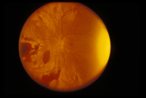
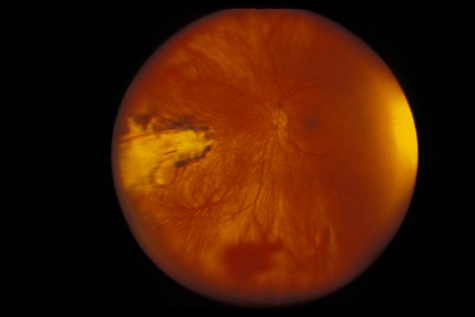
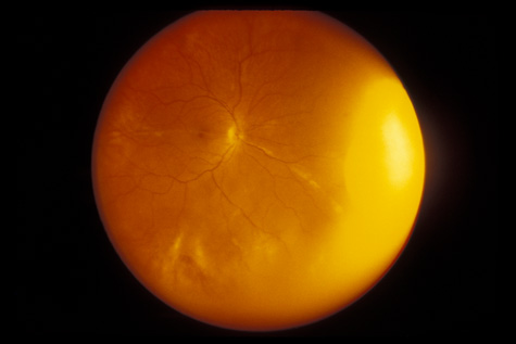
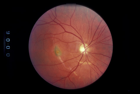
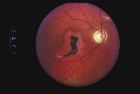
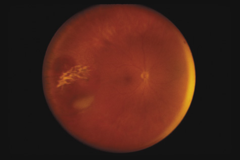
mechanism, or by ocular compression. COUP AND CONTRECOUP Courville24,25 introduced the concept of coup and contrecoup injury to explain brain damage caused by blunt trauma to the head. Coup refers to local trauma at the site of impact. Contrecoup refers to injuries at the opposite side of the skull caused by shock waves that traverse the brain. Foci of brain damage are found along the path of the shock waves, especially at interfaces of tissues of different density. The greatest difference in density is between the brain and the skull, and it is here that the most severe damage occurs. Wolter later used these concepts to explain eye injuries.26 Examples of coup injuries are corneal abrasions, subconjunctival hemorrhages, choroidal hemorrhages, and retinal necrosis (Fig. 1). The best example of a contrecoup injury is commotio retinae (Fig. 2). These injuries are discussed later in this chapter.
ANTERIOR–POSTERIOR COMPRESSION AND EQUATORIAL EXPANSION The volume of a closed space cannot be changed. Therefore, when the eye is compressed along its anterior–posterior axis, it must either expand in its equatorial plane (Fig. 3) or rupture. Using high-speed photography, Delori and associates27 studied blunt trauma in enucleated pig eyes. The cornea was indented 8.5 mm, reducing the original anterior–posterior axis by 41% and bringing the posterior surface of the cornea into contact with the iris and the lens. The equatorial plane was expanded to 128% of its original length. Specific examples of damage caused by this severe stretching of the ocular tissues are discussed later.
|
ANTERIOR SEGMENT INJURIES | ||||
CORNEA Abrasions of the corneal epithelium are common but because they heal rapidly, they are of little consequence. Endothelial damage can be more serious. A local concussion (coup effect) can rupture endothelial cells and loosen the intercellular tight junctions. They can also be damaged by being crushed against the iris and the lens. The transient corneal edema usually clears, but endothelial damage can be permanent. Follow-up studies of patients with traumatic hyphema show endothelial cell loss that correlates with the severity of the initial injury.28,29 When a total hyphema is present, the damaged corneal endothelium may permit hemoglobin to enter the corneal stroma. Usually, high intraocular pressure is required to cause corneal bloodstaining, but cases have been reported in which the intraocular pressure was normal.30 Corneal transplantation is rarely necessary as the bloodstaining nearly always clears spontaneously, but this may require a considerable period of time. The central cornea clears last. Adults can wait for clearing, but in young children, amblyopia can develop. When the cornea is abruptly forced backward by severe blunt trauma, it presses the iris against the lens, preventing the escape of aqueous into the posterior chamber. If enough force is applied, the entrapped aqueous dissects into the ciliary body, resulting in a recessed angle (Fig. 4). This tearing of the ciliary body is responsible for approximately 90% of the hyphemas seen after blunt trauma.31,32 Other causes of hemorrhage include separation of the iris from the ciliary body (iridodialysis) and sphincter tears. Ultrasound biomicroscopy can be useful in diagnosing dissection of the ciliary body or iridodialysis (Fig. 5).33–35
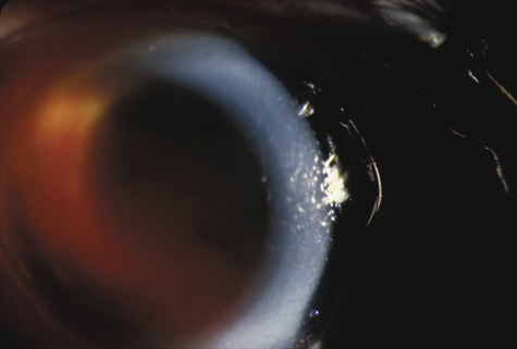
In injuries severe enough to cause a full-thickness corneal rupture, the wound usually crosses the limbus into the adjacent sclera. Exceptions are eyes that have had radial keratotomy, cataract surgery, or corneal transplantation, even years before the trauma.36–38 GLAUCOMA Acutely, the intraocular pressure may be normal, increased, or decreased. Aqueous outflow may be decreased because of angle recession, inflammatory trabeculitis, or hyphema. On the other hand, ciliary body injury tends to decrease aqueous inflow. The intraocular pressure depends on the balance of these factors. In patients who have large hyphemas, the pressure may rise abruptly. Therefore, it must be checked at regular intervals. It is especially important to monitor the intraocular pressure in hyphema patients who have sickle cell disease, be it homozygous or heterozygous. Unlike normal erythrocytes, sickled erythrocytes cannot pass through the trabecular meshwork and tend to occlude it, raising intraocular pressure. This, in turn, can cause central retinal artery obstruction even if the pressure is only in the high twenties or low thirties. Prompt paracentesis followed by surgical evacuation can restore vision.39 Late glaucoma can also develop, even years after the initial injury. Autopsy studies have found three ways by which the trabecular meshwork can be obstructed: proliferation of corneal endothelial cells and Descemet's membrane, proliferation of fibroblasts, and peripheral anterior synechiae (presumably from inflammation or from organization of blood in the angle for more than 7 days).40 Other possibilities are direct damage to the trabecular meshwork and damage to the ciliary muscle with reduced traction on the scleral spur. The risk is greatest in patients who have severe anterior segment damage such as cataract, angle recession, iris damage and displacement of the lens.41 Nine percent of patients with angle recession develop permanent glaucoma.42 If trabeculectomy is required, some authors recommend antimetabolites because the failure rate in patients with angle recession is greater than it is in patients with typical open angle glaucoma.43 LENS When the lens is struck by the cornea or by a strong shock wave, a transient anterior subcapsular cataract, known as a rosette cataract, may develop (Fig. 6). Repeated trauma, as in boxers, often causes posterior subcapsular cataract. Blunt trauma can also result in rupture of the anterior or posterior capsule.44,45 In severe cases, iridodonesis or a bead of vitreous in the anterior chamber signals a subluxed lens. Dislocation may also occur (Fig. 7). It is important to remember that ocular trauma is common, but lens dislocation is rare. Therefore, in patients with a dislocated lens, the clinician should always rule out predisposing causes such as Marfan's syndrome, homocystinuria and syphilis. In rare cases the lens itself can rupture and cause phacolytic glaucoma.
Blunt trauma can also result in transient high myopia caused by anterior shift of the lens-iris diaphragm secondary to ciliochoroidal effusion and ciliary body edema.46,47 UVEA Blunt trauma can lead to persistent uveitis. If traumatic uveitis is suspected, the ophthalmologist must carefully examine the fundus for posterior signs of trauma.48 HYPHEMA The management of traumatic hyphema remains somewhat controversial. The goals are to prevent peripheral anterior synechiae, severe acute glaucoma, and corneal blood staining. Although these can be the result of the initial hyphema, the risk increases dramatically with secondary hemorrhage which is most likely to occur 1 to 2 days after the injury. The risk is greatest in patients who present with large hyphemas and in African Americans, particularly if there is a history of sickle cell disease.49 The reported incidence of secondary hemorrhage ranges from 0% to 38%.39 Medical Management Currently, at the Wills Eye Hospital, most microhyphemas and many hyphemas are treated on an outpatient basis. It is appropriate to consider hospitalization for patients who are noncompliant, are at high risk of rebleeding, or have other severe injuries. Hospitalization can also be considered for children if child abuse is suspected or aggressive treatment is advised to prevent amblyopia. If treated as an outpatient, hyphema patients follow-up daily for 3 days. Microhyphema patients follow-up on the third day after initial trauma.50,51 The authors have long abandoned traditional strict bed rest but still encourage limited activity. The head of the bed should be elevated to encourage the blood to collect inferiorly, out of the visual axis. Eye-patching is avoided so that patients can immediately report decreased vision, which is often the first symptom of a secondary hemorrhage. The eye is protected with an eye shield. The authors dilate the pupil with atropine to reduce pain and to facilitate later fundus examination. Atropine may also reduce the risk of secondary hemorrhage by stabilizing the blood–ocular barrier. Topical corticosteroids are used to increase comfort and decrease inflammation.52–54 It has been reported that systemic corticosteroids markedly reduce the incidence of secondary hemorrhages,52–54 but two prospective double-blind studies found no beneficial effects.55,56 Aspirin and other nonsteroidal antiinflammatory agents must be strictly avoided because their antiplatelet effects predispose to rebleeding. Antifibrinolytic Agents Patients who are hospitalized, particularly for rebleeding, are treated with aminocaproic acid, which improves clot stabilization. Another antifibrinolytic agent is tranexamic acid. Antifibrinolytic agents prevent conversion of plasminogen to plasmin and block the action of plasmin itself, theoretically preventing retraction of clots that occlude injured blood vessels until they are more completely repaired. At the Wills Eye Hospital, it reduced secondary hyphema from 25% to 10%. Several studies showed that aminocaproic acid reduces the incidence of secondary hyphema from approximately 30% to approximately 4%.57–59 Caution is necessary when antifibrinolytic agents are discontinued, because in some patients, the clot may rapidly dissolve 1 or 2 days later with a resultant increase in intraocular pressure severe enough to require surgical intervention.60 To try to avoid this complication, the authors decrease the dose of aminocaproic acid by one-half after 48 hours. Patients are discharged after 72 hours and reexamined daily for a few more days. Some studies of topical aminocaproic acid suggest that it may be effective in reducing incidence of rebleeding,61,62 although this is controversial.63 Management of Intraocular Pressure It is important to keep the intraocular pressure under control to prevent corneal bloodstaining. If it is elevated, the authors initially treat with β-blockers, followed by an α-agonist (e.g., apraclonidine 0.5% or brimonidine 0.2%) or carbonic anhydrase inhibitor (e.g., dorzolamide 2% or brinzolamide 1%). Prostaglandin analogues and miotics are avoided because these may increase inflammation. If these topical medications are not adequate, oral acetazolamide, (Diamox®) is used, followed by mannitol if needed. If mannitol is necessary to control the intraocular pressure surgical evacuation may be indicated. When treating patients with sickle disease, it is preferable to avoid treatments that could promote sickling. For example, carbonic anhydrase inhibitors may promote sickling by increasing aqueous levels of ascorbic acid. Adrenergic agonists with predominantly α1 effects should also be avoided. It is also best to avoid repeated use of hyperosmotic agents such as mannitol, which results in hemoconcentration and greater blood viscosity.39 Instead of Diamox (Goldshield Pharmaceuticals, Surrey, UK), methazolamide (Neptazane, Lederle Pharmaceutical Division, American Cyanamid Company, Pearl River, NY ) can be tried as it has less acidifying effect. The authors continue treatment until intraocular pressure is less than 30 mm Hg (24 mm Hg for patients with sickle cell disease). Recently, transcorneal oxygen therapy has been attempted with encouraging results for decreasing intraocular pressure. By increasing the partial pressure of oxygen, cells in the anterior chamber are less likely to sickle.64 Indications for Surgery Surgery for hyphema is discussed in detail elsewhere in these volumes. The most common indications are corneal blood staining, total hyphema for 1 to 2 days, large clots present for 5 to 6 days, and uncontrolled intraocular pressure. In patients with sickle cell trait or disease evacuation is strongly considered if the patient has a marked decrease in vision or intraocular pressure above 24 mm Hg. Combining a trabeculectomy with the washout in patients with high intraocular pressure has been advocated.65 |
POSTERIOR SEGMENT INJURIES | ||||||||||||||||
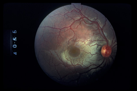
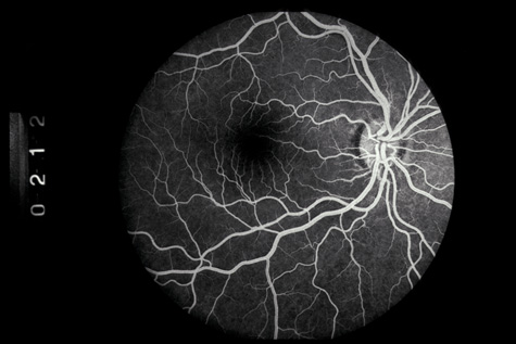
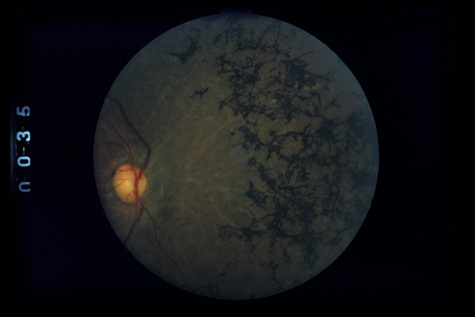
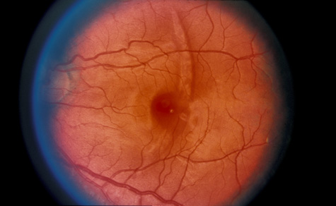
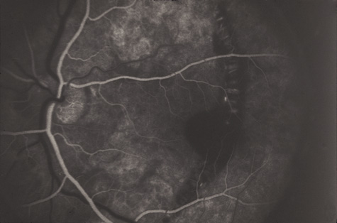
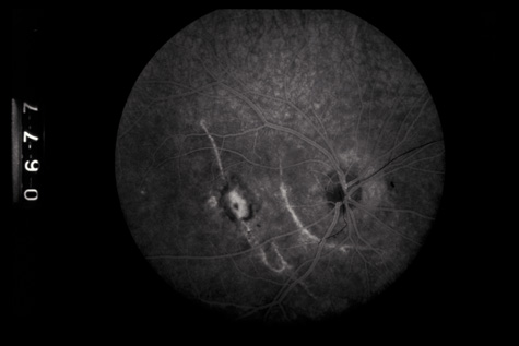
COMMOTIO RETINAE Commotio retinae (Latin, meaning retinal contusion) is a contrecoup injury. It can occur peripherally (Fig. 8) or centrally, in which case it is called Berlin's edema (Fig. 9). Immediately and for several hours after the trauma, the retina appears normal, although the patient may complain of decreased vision. Thereafter, the outer layers of affected retina become opaque. On fluorescein angiography, the opaque retina blocks background choroidal fluorescence, and in most cases there is no leakage into or under the retina (Fig. 10). For years, clinicians had difficulty explaining this blockage, because leakage is expected in conditions with edema. It was then shown in experimental animals and in human autopsy eyes that Berlin's edema is not true edema. The retinal opaqueness is the result of intracellular edema and fragmentation of the photoreceptor outer segments and intracellular edema of the underlying pigment epithelium. There is little or no intercellular fluid.66–69
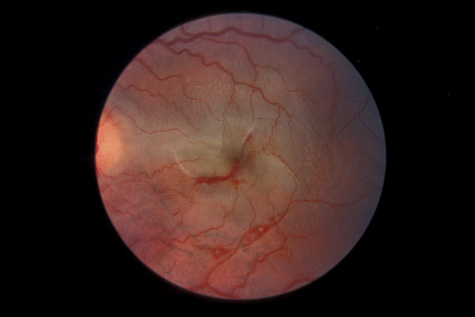
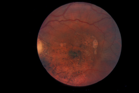
The visual acuity in commotio retinae varies from 20/20 to 20/400 and does not always correlate with the degree of retinal opacification. There is no known treatment. The prognosis is usually excellent except in cases with associated subfoveolar choroidal rupture and in cases with choroidal rupture with subfoveolar hemorrhage. Poor visual recovery can also be expected in cases with severe retinal pigment epithelial damage. Serous retinal detachment (Fig. 11) signals this condition, which can be confirmed by leakage of fluorescein into the subretinal space.70 The late manifestations of contrecoup injury to the retinal pigment epithelium (RPE) vary from minor atrophic changes that are seen as transmission defects on fluorescein angiography to massive hyperplasia and migration of the RPE. This later condition results in bone corpuscular and granular pigmentation that resembles retinitis pigmentosa (Fig. 12).71,72 The traumatic pigmentary changes may be confined to the posterior pole, to the periphery, or to certain quadrants, or they may be more widespread. When the trauma to the RPE destroys photoreceptor cells, localized visual field defects result. Arcuate field defects are not found because the overlying nerve fiber nearly always remains intact. MACULAR HOLES In rare cases the retinal contusion causes cystoid macular edema that may, in turn, progress to a macular hole (Fig. 13). A macular hole can also be caused by acute posterior vitreous detachment with foveal traction. In such cases, an overlying operculum is seen. Surgically, the macular hole can sometimes be closed with vitrectomy and gas injection.73 However, in some cases macular holes can spontaneously close, and because of this possibility some suggest observation for 4 to 6 months after the injury.74,75
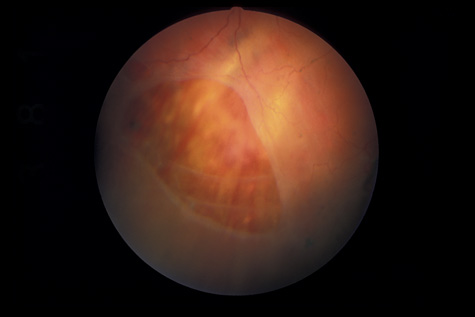
RETINAL TEARS Necrotic Tears If a blunt object strikes the eye posterior to the ora serrata, the direct concussive effect on the retina (coup effect) may cause full-thickness retinal necrosis with subsequent retinal detachment (Fig. 14). The underlying RPE is usually damaged as well.
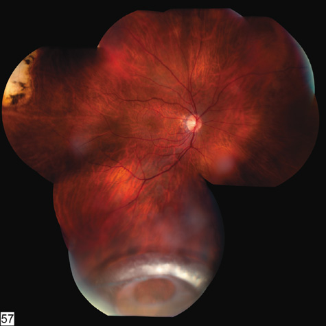
Vitreous Traction Tears Weidenthal and Schepens76 concluded, from their study of trauma to enucleated pig eyes, that rapid equatorial expansion is responsible for tears at the anterior and posterior borders of the vitreous base. Because the vitreous body is relatively elastic, slow compression of the eye is not deleterious. However, when the eye is rapidly compressed, the vitreous does not have enough time to stretch. As a consequence, there is strong traction at the vitreous base that may cause tears at its anterior and posterior borders. Scott suggested an alternative mechanism for these tears (J. Scott, personal communication, 1982). He suggested that retrodisplacement of the cornea and aqueous drives the lens-iris diaphragm backward against the anterior vitreous, forcing the vitreous back and pushing the vitreous base away from its adherence to the pars plana epithelium and anterior retina. Whatever the mechanism, a portion of the vitreous base can be avulsed from the retina and pars plana. The avulsed base looks like a ribbon floating in the vitreous cavity. Closer inspection reveals that the vitreous base remains in contact with the adjacent vitreous cortex, which is also avulsed (Fig. 15). Avulsion of the vitreous base is pathognomonic of blunt trauma and may have considerable medicolegal importance. Unfortunately, in most cases of severe trauma, the vitreous base does not separate cleanly from the retina and pars plana epithelium. It remains adherent, tearing these tissues. The retina can be torn along the posterior margin of the vitreous base, or the nonpigmented pars plana epithelium can be torn along the anterior margin of the vitreous base, or both can be torn simultaneously. Similarly, if the vitreous is strongly adherent to either lattice degeneration or a vitreoretinal scar posterior to the vitreous base, a posterior flap tear may occur.77 Any of these tears can cause a retinal detachment.
Tears along the anterior and posterior margins of the vitreous base are most common inferotemporally. The next most common location is superotemporal. When located superonasally, they are nearly always caused by trauma.78 As regards true dialyses, some authors feel that all dialyses, even inferotemporal ones, are traumatic.79 However, many are familial, bilateral, or found in patients with no historical or histopathologic evidence of injury.80 In these cases, there is probably a developmental abnormality of the inferotemporal peripheral retina and vitreous base. Tasman's clinical study81 confirmed Weidenthal's experimental evidence that tears caused by blunt trauma nearly always occur at the time of injury. He prospectively examined 52 patients with hyphema. Nine had an acute dialysis, but in 2 years of follow-up, only one late break was found, and that was in a patient whose initial hemorrhage had prevented complete examination of the periphery. Other investigators have confirmed this conclusion.82 Stretch Tears Occasionally, rapid horizontal expansion of the eye can produce a stretch tear of the retina (Fig. 16). These curvilinear breaks are usually concentric to the optic nerve. They can cause retinal detachment, but in some cases, they are self-sealing (Fig. 17). Glial cells migrate across the undetached posterior cortical vitreous, form a membrane, and then contract, closing or partially closing the breaks and preventing detachment.
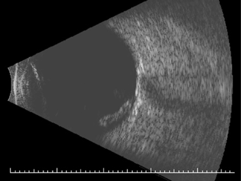
The Role of Head Trauma in Retinal Tears Ophthalmologists have long argued about whether or not trauma to the head but not directly to the eye or orbit can cause a retinal tear. Doden and Stark83 showed that it probably does not, at least in normal eyes. He examined 247 patients who had received a severe blow to the head, and none had a retinal tear. However, the authors have seen acute posterior vitreous detachment (PVD) and retinal tears immediately after a blow to the head in patients who were predisposed to retinal tears because of high myopia, lattice degeneration, or other abnormalities RETINAL AND VITREOUS HEMORRHAGE If the retina is torn, blood vessels that bridge the tears may bleed into the vitreous. Vitreous hemorrhage can also result from acute PVD and avulsion of superficial retinal vessels. A third possible mechanism is rupture of the ciliary body. In many cases the source of the bleeding is never found. Ultrasound examination is indicated if the posterior segment is obscured by hemorrhage. Ultrasound can reveal a retinal tear or detachment, choroidal detachment, posterior vitreous detachment, or occult ruptured globe. Bed rest with head elevation encourages the blood to settle and improves the view to the fundus on follow-up examinations. RETINAL DETACHMENT Because men and boys are most likely to be engaged in fighting or contact sports, it is not surprising that at least three-fourths of the patients with traumatic retinal detachment are men or boys,84 and that blunt trauma is the leading cause of retinal detachment in children and adolescents.85 Because the affected patients have a formed vitreous, traumatic retinal detachments typically progress slowly unless a giant tear is present. In some patients, the trauma occurs months or years before the detachment is diagnosed. Demarcation lines, atrophy of the underlying pigment epithelium, subretinal precipitates, retinal macrocysts, and extensive vitreous “tobacco dust” are all commonly seen (Fig. 18). Proliferative vitreoretinopathy is uncommon, so the prognosis for reattachment is excellent, provided, of course, that all breaks are found. In 87% of traumatic retinal detachments, the causative tear is found at the vitreous base. Superonasal breaks at the anterior vitreous base are commonly overlooked. Because many traumatic retinal breaks cause subsequent retinal detachment, they should all be treated with laser or cryotherapy.
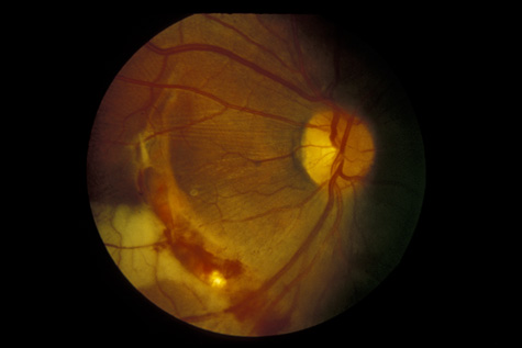
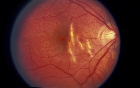
CHOROIDAL INJURIES Posterior choroidal ruptures are probably caused by anterior-posterior compression and equatorial expansion. The retina is relatively elastic, and the sclera is relatively tough. Both resist ruptures. Bruch's membrane, on the other hand, is inelastic and more prone to rupture. The overlying RPE and underlying choriocapillaris are also torn, but in most cases, the deep choroidal blood vessels remain intact (Fig. 19). Rupture of the choriocapillaris often results in subretinal hemorrhage. Patients with angioid streaks and other conditions known to be associated with a inelastic and fragile Bruch's membrane are especially vulnerable to choroidal rupture (Fig. 20).
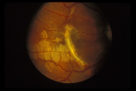
Initially, a choroidal rupture may be obscured by a subretinal hemorrhage caused by tearing of the choriocapillaris. Later, after the blood has resorbed, a white curvilinear streak concentric to the optic disc is seen. Only rarely is a rupture oriented radially with respect to the optic disk. Most are temporal to the disc and single, although nasal and multiple ruptures can also occur (Fig. 21).
If the choroidal rupture is under the foveola or if an associated subretinal hemorrhage extends under the foveola, the visual prognosis is generally poor (Fig. 22); however, a recent study showed that even patients with foveal or multiple choroidal ruptures can regain good central vision after extended follow-up.86 If the rupture is elsewhere the prognosis is good because the overlying nerve fiber layer is almost never torn. Therefore, a rupture can be located between the disc and the macula and yet not affect the visual acuity (Fig. 21).
Similar to many conditions that damage Bruch's membrane, choroidal ruptures can, months to years later, be complicated by development of a choroidal neovascular membrane, with serous or hemorrhagic retinal detachment and loss of central vision (Fig. 23).87–89 They usually respond well to laser therapy. Also, studies have demonstrated that subfoveal choroidal neovascular membranes from etiologies other than age-related macular degeneration can benefit from photodynamic therapy.90,91
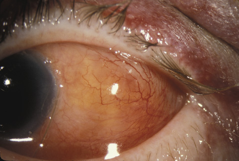
SCLERAL RUPTURE In eyes that have not undergone prior surgery, the two most common locations for scleral rupture are at the limbus (under intact conjunctiva) and parallel to the muscle insertions between the insertion and the equator. Radial and posterior ruptures are relatively uncommon.29 The hallmarks of scleral rupture are severe reduction in visual acuity, an afferent pupillary defect, hypotony (although a normal intraocular pressure does not rule out a small rupture), an abnormally deep anterior chamber, decreased ocular ductions, severe subconjunctival edema (Fig. 24), hyphema, and vitreous hemorrhage.92 The diagnosis can rarely be confirmed by ophthalmoscopy because severe vitreous hemorrhage, hyphema, or both nearly always accompany scleral rupture. Ultrasonography and computed tomography (CT) scanning may be helpful. Both show a shrunken globe. In addition, the CT scan shows subconjunctival edema (Fig. 25). In eyes in which the anterior chamber depth cannot be seen because hyphema, ultrasonography and CT scanning often show a deepened chamber. CT scanning is also useful in identifying any intraocular foreign bodies.
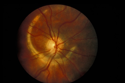
The prognosis for recovery of useful vision in a ruptured globe is very poor, even with current vitrectomy techniques, because the underlying choroid and retina are also torn. Fibrovascular tissue proliferates into the eye and causes severe massive periretinal proliferation. Although most eyes usually only have one rupture, the authors and others93 have seen eyes with more than one. OPTIC NERVE INJURIES The optic nerve can be severely damaged by blunt trauma, although the precise mechanism is not known. Possible mechanisms include compression by intrasheath hemorrhage and edema and direct shock-wave trauma to the nerve fibers. The management of traumatic optic neuropathy is controversial. The only prospective, controlled, randomized trial, the International Optic Nerve Trauma Study, failed because of inadequate recruitment.94 Many authorities advocate high-dose intravenous steroids, especially if they can be started within 8 hours. Other treatments that are based on CT findings include surgical decompression of optic nerve sheath hematoma and neurosurgical decompression of the optic canal.95 However, two recent studies did not show that any of these treatments improved visual outcome.94,96 Another severe complication of blunt trauma is evulsion of the optic nerve (Fig. 26). Although this usually is accompanied by severe damage to other ocular tissues, it can be the sole manifestation of apparently minor direct trauma or even a blow to the occiput.
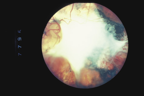
RETINITIS SCLOPETERIA Retinitis sclopeteria is the rupture of the choroid or retina caused by shock waves generated by passage of a high-velocity missile through the orbit without directly striking the eye. Initially a subretinal or vitreous hemorrhage is seen. If the optic nerve is damaged, visual acuity can be profoundly decreased. In severe cases, massive amounts of fibrous tissue proliferate into the eye (Fig. 27). In others, as the blood clears, a claw-like break is often seen in Bruch's membrane and in the choriocapillaris (Fig. 28). Retinal detachment rarely occurs at the site of the injury, probably because of binding of the retina to the choroid by fibrous tissue, but late detachment from a break at a distal site can occur.97,98
|