Chapter 34
Phakomatoses
PETER V. PALENA and JAMES J. AUGSBURGER
Main Menu   Table Of Contents

Search

TUBEROUS SCLEROSIS (BOURNEVILLE'S DISEASE)
NEUROFIBROMATOSIS
VON HIPPEL-LINDAU SYNDROME
STURGE-WEBER SYNDROME
RETINAL-NEURO-CUTANEOUS CAVERNOUS HEMANGIOMA SYNDROME (WESKAMP-COTLIER SYNDROME)
WYBURN-MASON SYNDROME
REFERENCES

The phakomatoses are a group of clinical disorders that are linked, at least in a historical sense, by various attributes of the component lesions, the organs involved, and the pattern of clinical inheritance observed in some cases. Although the term phakomatoses was coined by van der Hoeve in 1923 in a paper concerning the similarities between von Recklinghausen's neurofibromatosis and Bourneville's tuberous sclerosis,1 this term has never been satisfactorily defined. Absolute inclusion criteria for classifying a disorder as a phakomatosis were not presented by van der Hoeve, and a consensus about such criteria has not been reached in the intervening years.2

In the absence of a completely satisfactory, generally agreed upon definition of the term phakomatosis, confusion reigns and will probably continue to reign for the foreseeable future. Some authors believe that all phakomatoses are neuro-oculocutaneous syndromes with autosomal dominant inheritance. Others believe that one or more characteristic skin lesions must be present in a substantial number of patients with a neuro-ocular syndrome to warrant its classification as a phakomatosis. Still others believe that the essential element of a phakomatosis is the presence or development of multiorgan hamartomas.3

For the purposes of this chapter, we will define the phakomatoses as a group of independent clinical syndromes characterized by multiple tumors, some of which are or can become malignant, arising in disparate organs of the body. These syndromes are important to ophthalmologists because they all have ophthalmic tumors as prominent clinical manifestations in at least some affected individuals. It is the patients with ophthalmic lesions as part of a multiorgan tumor syndrome that are the particular subject of this chapter.

Three syndromes are consistently classified as phakomatoses by most authors and also meet our definitional criteria: tuberous sclerosis (Bourneville's disease), neurofibromatosis, and the von Hippel-Lindau syndrome. The Sturge-Weber syndrome (and its variants) is commonly classified as a phakomatosis but does not precisely conform to our definition. Several other syndromes, including the Wyburn-Mason syndrome, the retinal-neurocutaneous cavernous hemangioma syndrome described by Weskamp and Cotlier, and others, are also classified as phakomatoses by some authors but do not conform to our definition.

Back to Top
TUBEROUS SCLEROSIS (BOURNEVILLE'S DISEASE)
Tuberous sclerosis is a multiorgan tumor syndrome with prominent ophthalmic manifestations.4 The complete syndrome is characterized by multifocal, bilateral retinal astrocytic hamartomas, several unusual cutaneous lesions, astrocytic tumors of the brain, mental retardation, seizures, and a variety of cysts and tumors of other organs.5 The clinical spectrum of this syndrome is extremely broad, ranging from minimal to marked in affected individuals. Many persons with limited forms of the disease are probably not recognized as having tuberous sclerosis.

The prevalence of tuberous sclerosis in the general population is at least 1 in 10,000.6 About one third of cases are familial, whereas two thirds are sporadic. There is no recognized racial predilection, and the sexes are affected equally. Signs and symptoms of tuberous sclerosis usually begin before the patient is 6 years old.

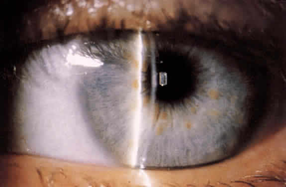
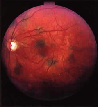
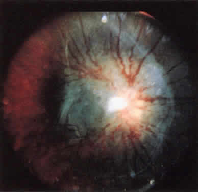
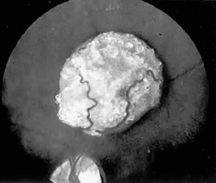
The classic ophthalmoscopic feature of tuberous sclerosis is the retinal astrocytoma (astrocytic hamartoma).7 This lesion arises within the nerve fiber layer of the retina. It appears more frequently in the posterior fundus than in the periphery. Small lesions commonly appear as translucent intraretinal patches with minimal thickness. Slightly larger lesions usually appear more opalescent or even opaque white. Large lesions tend to be opaque white, substantially elevated, and frequently multilobulated (Fig. 1). Intralesional calcification develops within some larger lesions. Multiple lesions can be present in one eye, and many affected patients have binocular involvement. The retinal blood vessels associated with these lesions tend not to be dilated or tortuous in most patients. If lesion growth occurs, it tends to be extremely slow. Approximately one half of patients with tuberous sclerosis have at least one typical astrocytic retinal hamartoma.8

Fig. 1. Astrocytic hamartoma of the retina in tuberous sclerosis.

Lesions similar to those that occur in the retina commonly develop in the cerebrum and less commonly develop in the basal ganglion, brain stem, and cerebellum. Both retinal and central nervous system (CNS) lesions can be calcified and identified radiologically.9 Epilepsy and mental deficiency are common in severely affected individuals, and early death occurs in occasional patients as a result of progressive CNS tumor expansion or status epilepticus.5

A variety of unusual tumors develop in the heart, kidney, lungs, thyroid, and other visceral organs in some patients with tuberous sclerosis.5 The classic cardiac tumor is the rhabdomyoma, which can lead to conduction abnormalities, heart failure, and premature death. Angiomyofibromas tend to develop in the kidney, and these lesions can lead to death from renal failure.

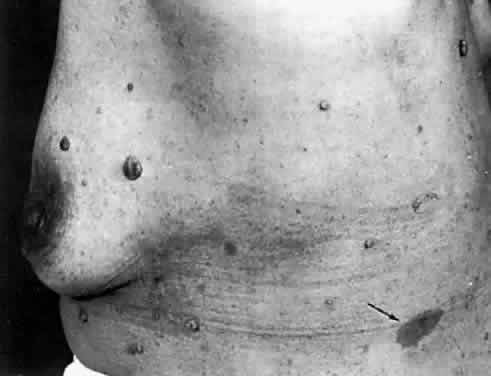
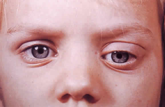
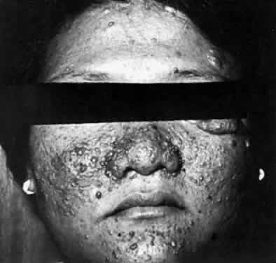
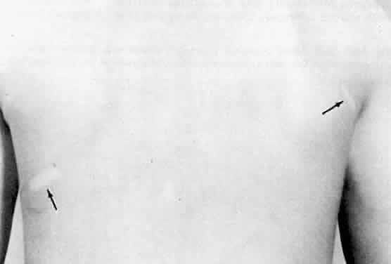
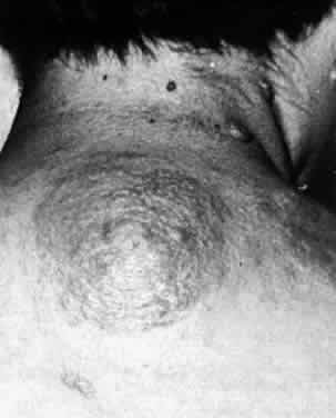
Several cutaneous lesions are prominent features of tuberous sclerosis. Adenoma sebaceum is an unusual facial dermatologic eruption characterized by pinhead- to pea-sized yellowish to reddish-brown papules distributed in butterfly fashion over the nose, cheeks, and nasolabial folds (Fig. 2). Histopathologically, the individual skin lesions are angiofibromas.10 Ash leaf spots are congenital white or hypomelanotic skin macules ranging from about 1 mm to several centimeters in diameter and having a configuration resembling an ash leaf (Fig. 3). These lesions are most easily demonstrated under ultraviolet light (Wood's lamp).11 The shagreen patch is a thickened patch of skin with the texture of pigskin or sharkskin, usually occurring over the lower back (Fig. 4). Subungual fibromas are benign fibrous tumors that develop at the sides of the nail beds in some patients.

Fig. 2. Butterfly distribution of papular angiofibromas of adenoma sebaceum in tuberous sclerosis.

Fig. 3. Hypomelanotic macules (arrows), the ash leaf lesions in tuberous sclerosis. (Courtesy of Dr. DL Baxter.)

Fig. 4. Pigskin or orange-peel appearance of shagreen patch in tuberous sclerosis.

Treatment of affected individuals is purely symptomatic at present. Death tends to occur in many severely affected individuals by the third decade as a result of the CNS lesions or visceral tumors.

Recent molecular biologic studies have identified loci on the long arm of chromosome 9 (9q32-34), on the long arm of chromosome 11, on the short arm of chromosome 16 (16p13.3), and on the long arm of chromosome 12 (12q22-24.1) as tuberous sclerosis genes in different multigenerational families that have been studied.6 Of these loci, the 9q32-34 locus has been the most consistent, being associated with one third to one half of all familial cases.

Systemic evaluation of individuals suspected of having tuberous sclerosis should probably include fundus examination, dermatologic evaluation to identify characteristic skin lesions, magnetic resonance imaging (MRI) of the central nervous system,12 and computed tomography (CT) or MRI of the abdominal viscera. Examination of family members to look for a familial pattern is also appropriate.

Back to Top
NEUROFIBROMATOSIS
Neurofibromatosis (NF) is a term that has been applied to a variety of related syndromes, all of which are characterized by neuroectodermal tumors arising within multiple organs and autosomal dominant inheritance. Two distinct clinical forms of neurofibromatosis are currently recognized.

NF-1 (peripheral neurofibromatosis, classic neurofibromatosis, von Recklinghausen's disease13) is characterized by café au lait spots, axillary and inguinal freckling, Lisch nodules of the iris, several types of cutaneous neurofibromas, optic nerve gliomas, and neurofibromas of the central nervous system.14 The café au lait spots in this syndrome tend to be more numerous and larger than the lesions arising nonsyndromically in the general population. Six or more café au lait spots larger than 1.5 cm in diameter are generally considered diagnostic of NF-1.15 Axillary and inguinal freckling are present in about 90% to 95% of affected individuals.16 Lisch nodules, which are melanocytic hamartomas of the iris stroma,17 appear as tan to light brown nodules studding the surface of the iris (Fig. 5). These lesions are rarely if ever present at birth but tend to develop by the second to third decades of life in almost all persons with NF-1.17 Subcutaneous neurofibromas in NF-1 tend to be either pedunculated tumors, which tend to develop in various regions of the body (Fig. 6) and are commonly found on the eyelids, or diffuse plexiform neuromas, which have the texture of a “bag of worms” on palpation.14 The optic nerve gliomas in NF-1 tend to cause proptosis (Fig. 7) and optic atrophy in early childhood.18 They are sometimes bilateral and occasionally arise from the optic chiasm or tracts instead of the optic nerves proper. CNS neurofibromas can cause hemiparesis, hemiatrophy, and seizures in some affected individuals.14

Fig. 5. Lisch nodules of the iris in neurofibromatosis type 1 (NF-1).

Fig. 6. Isolated neurofibromas, some of which are umbilicated, are seen over the trunk. Typical café au lait spot also present (arrow). (Courtesy of Dr. DL Baxter.)

Fig. 7. Proptosis due to orbital optic nerve glioma in youngster with NF-1.

NF-2 (central neurofibromatosis) is typified by bilateral acoustic neuromas and widely scattered neurofibromas, meningiomas, gliomas, and schwannomas.19 Café au lait spots, axillary freckling, and Lisch nodules are uncommon in patients with NF-2. The most consistent problem suffered by patients with NF-2 is sensorineural deafness due to the acoustic nerve tumors.

Neurofibromatosis appears to be the most common of the phakomatoses, affecting approximately 1 person per 3500.14 NF-1 appears to account for the majority of cases. The frequency of NF-1 has been estimated to be approximately 1 in 3500 to 4000,20 but that of NF-2 appears to be no greater than 1 in 50,000. Males and females appear to be affected with equal frequency, and there does not appear to be any racial predilection for the syndrome.

Although some of the features of NF can be apparent congenitally, most of the signs and symptoms of these syndromes do not become apparent until late childhood or early adulthood. The severity of the syndrome varies markedly from patient to patient. Consequently, many patients with limited forms of NF are probably not currently diagnosed.

Ophthalmologic findings in NF-1 include the Lisch nodules of the iris (present in over 95% of adult patients)17 and subcutaneous pedunculated and plexiform neurofibromas of the eyelids, as mentioned above, as well as occasional retinal tumors indistinguishable from the retinal astrocytic hamartomas found in tuberous sclerosis.21,22 Glioma of the optic nerve is present in approximately 10% to 15% of affected patients.23 Proptosis can occur as a result of an optic nerve glioma or an orbital neurofibroma, and pulsating exophthalmos or enophthalmos due to a congenital anomaly of development of the sphenoid bone is also a feature of some cases of NF-1.24 Congenital or infantile glaucoma appears to be more common in patients with this syndrome.21 Some affected patients develop multifocal bilateral melanotic fundus lesions as a feature of the syndrome (Fig. 8).22,24

Fig. 8. Multifocal melanotic fundus lesions in an adult patient with NF-1.

Ophthalmologic findings in NF-2 are uncommon.25 Lisch nodules of the iris, eyelid neurofibromas, and optic nerve gliomas are not generally present in patients with this syndrome. In recent years, however, several young patients with NF-2 have been found to have a combined hamartoma of the retina (Fig. 9) in one or both eyes.26 If the lesion involves the macula, the vision tends to be poor.

Fig. 9. Presumed combined hamartoma of neurosensory retina and retinal pigment epithelium in a child with neurofibromatosis type 2 (NF-2).

Symptoms in patients with NF-1 and NF-2 depend on the location and extent of the tumors. Treatment, when necessary, generally consists of surgical excision of symptomatic tumors, if that is possible. External-beam radiation therapy has been used to treat some gliomas of the optic nerve that are not amenable to surgical excision,27 but the effectiveness of such treatment has not been confirmed in comparative clinical trials.

Molecular biologic investigations have identified distinct chromosomal abnormalities in NF-1 and NF-2. A gene that codes for NF-1 has been localized to the long arm of chromosome 17 at locus 17q11.2,20 and a totally different gene on the long arm of chromosome 22 at locus 22q12 has been linked with NF-2.28

Back to Top
VON HIPPEL-LINDAU SYNDROME
The von Hippel-Lindau syndrome (VHLS) is a multiorgan hamartomatosis characterized by retinal vascular hamartomas, similar vascular hamartomas of the central nervous system, a host of other solid and cystic hamartomas and hamartias, and malignant tumors, the most important of which are renal cell carcinoma and pheochromocytoma.29 The full-fledged syndrome commonly runs in families with a clear autosomal dominant inheritance pattern. Affected individuals are at substantial risk of early death, usually as a result of their intracranial hemangiomatous lesion or renal cell carcinoma.29

VHLS appears to be rare, but its precise incidence has never been determined.30 In patients with full-fledged VHLS, one or more clinically identifiable manifestations of the disease are usually present by or before the third decade of life.29

Not all patients who develop a hemangioblastoma of the retina or central nervous system have VHLS. In fact, most patients with just one of these lesions do not develop any other manifestations of VHLS and have no other affected family members. These individuals are considered to have a sporadic, nonsyndromic disorder. Most individuals of this type develop only a single lesion in one eye or a single hemangiomatous lesion of the central nervous system. If a patient has only the characteristic retinal lesion and its associated ocular complications (see below), then that patient is said to have “von Hippel's disease” (angiomatosis retinae)31 and not VHLS. Approximately 80% of patients with von Hippel's disease have a single retinal hemangioblastoma in one eye and no family history of the ocular disease. In contrast, about 20% of patients with von Hippel's disease have a positive family history of the ocular disorder, and most of these patients have bilateral, multifocal retinal hemangioblastomas and other features of VHLS.

The classic ocular lesion of von Hippel-Lindau disease is a globular red retinal tumor visible on ophthalmoscopy or slit lamp biomicroscopy of the ocular fundus. Such tumors have been shown by histopathological, immunohistochemical, and ultrastructural analysis to be benign retinal hemangioblastomas composed of endothelial-lined vascular channels in a stroma consisting of polyhedral, spindle-shaped, and vacuolated glial cells.32 Such tumors are commonly referred to as “capillary hemangiomas,” although this is not precisely correct from a pathologic point of view.

Individual retinal hemangioblastomas range from tiny red intraretinal dots too small to be reliably identified as hemangiomas by ophthalmoscopy, to large tumors (greater than 10 mm in diameter) filling a quadrant or more of the fundus. The typical retinal hemangioblastoma that is larger than about 0.5 mm in diameter is associated with dilated, tortuous afferent and efferent retinal blood vessels. The larger the retinal tumor, the more prominent these afferent and efferent retinal vessels tend to be. Most patients with complete VHLS develop multiple retinal hemangioblastomas in both eyes.29

Most retinal capillary hemangiomas that are clinically identifiable by ophthalmoscopy give rise to an exudative intraretinal and sometimes subretinal response. In pronounced cases, this can result in a massive accumulation of intraretinal and subretinal exudates. In fact, some eyes with one or more peripheral retinal capillary hemangiomas develop a mound of macular exudates (exaggerated macular exudative response) that markedly reduces vision in that eye. In occasional eyes, proliferative vitreoretinopathy develops in response to the retinal hemangiomas, and this phenomenon can lead to a partial or total tractional retinal detachment.

Retinal capillary hemangiomas can arise from all retinal sites, from the optic disc to the periphery. However, they tend to be more common in the fundus midzone to periphery and rarely develop in the macula. The retinal vascular tumors are bilateral, multifocal, or both in at least 50% of affected patients with the familial form of the disease.29

Progression of retinal capillary hemangiomas in VHLS, as in patients with sporadic von Hippel's disease, is highly variable, but tumor enlargement, hemorrhages, exudates, gliosis, and retinal detachment may develop, eventually resulting in glaucoma or phthisis bulbi.

The classic CNS lesions of VHLS are solid and cystic cerebellar hemangioblastomas.33,34 The component cells in these tumors appear quite similar to those in the retinal hemangioblastomas that have been studied.32 Similar vascular lesions also occur in some patients in the medulla and spinal cord. In addition to the retinal and CNS lesions, patients with von Hippel-Lindau syndrome tend to develop renal cell carcinoma (hypernephroma), pheochromocytoma, and occasionally islet cell carcinoma of the pancreas, any of which can prove fatal, as well as cystadenomas of the pancreas and epididymis, cysts in the kidneys, pancreas, and ovaries, and polycythemia.29,30 Although each of the three classic phakomatoses described earlier in this chapter has characteristic cutaneous lesions as a prominent feature, VHLS is not associated with any typical skin lesions.

Signs and symptoms of VHLS and the necessity for treatment depend on the nature of the lesion, the location and size of the lesions, and the symptoms they are producing. Most patients with VHLS who are initially identified on the basis of symptoms present with cerebellar symptoms or visual problems.29 Many other affected individuals in recognized VHLS families are identified on baseline screening studies or periodic follow-up examinations prior to the development of any symptoms.

Treatment of small to medium-sized retinal capillary hemangiomas generally consists of photocoagulation,35 cryotherapy,36 or both, whereas treatment of larger retinal lesions can entail more heroic measures, such as penetrating diathermy,37 episcleral plaque radiation therapy, charged particle beam irradiation,38 or even microsurgical resection.39 Unfortunately, visual improvement is unlikely in advanced cases that are complicated by total retinal detachment, and many such eyes are eventually enucleated because of pain or phthisis bulbi. Treatment of the CNS and visceral lesions of this disease is generally surgical and is beyond the scope of this chapter.

Molecular biologic studies in recent years have localized a genetic defect in VHLS to the short arm of chromosome 3 (locus 3p25-26).40 In affected kindreds, screening for the VHLS gene by DNA polymorphism analysis allows for detection of unaffected gene carriers and affected individuals who have subclinical lesions at the time of investigation.

Because of the frequency and severity of the various multiorgan lesions in VHLS, a comprehensive protocol of periodic re-examination and ancillary testing of affected patients in identified VHLS families is almost certainly appropriate.30 Some of the elements of an “optimal” protocol (assuming that sufficient financial resources and technology are available) include the following:

  1. Annual complete physical examination, including neurologic examination and comprehensive ophthalmoscopy of both eyes
  2. Annual urine testing for vanillylmandelic acid
  3. High-resolution magnetic resonance imaging of the brain every 3 years up to age 50 and every 5 years thereafter
  4. Annual renal ultrasound scan
  5. CT scan of the abdomen every 3 years (more frequently if multiple renal cysts are present)

An unresolved issue is the appropriate baseline investigation and subsequent follow-up evaluation of patients with a single retinal or CNS hemangioblastoma and no family history of VHLS. Aggressive baseline evaluation and follow-up are probably more appropriate for patients who present with their CNS or retinal hemangioblastoma early in life than for those with a lesion that is first detected at age 40 years or older.

Back to Top
STURGE-WEBER SYNDROME
The Sturge-Weber syndrome (SWS) is a dermato-oculoneural syndrome characterized by cutaneous facial nevus flammeus in the distribution of the branches of the trigeminal nerve, ipsilateral cavernous hemangiomas of the meninges and the choroid, and ipsilateral congenital, infantile, or juvenile glaucoma.41,42 The lesions in the eye, skin, and brain in SWS are always present at birth (i.e., they are birthmarks or congenital anomalies rather than true tumors like those in the three classic phakomatoses). Some patients with the full-fledged syndrome develop a seizure disorder that may be refractive to most conventional therapies, and a portion of affected individuals are mentally retarded.

The majority of patients with SWS have sporadic, nonfamilial disease. Only a few familial clusters of the syndrome have ever been reported, and most of those have not exhibited the clear-cut autosomal dominant inheritance pattern of the three classic syndromes. The incidence of the complete syndrome and its formes frustes is unknown.

The classic cutaneous feature of SWS is the facial nevus flammeus, a flat to moderately thick zone of dilated telangiectatic cutaneous capillaries lined by a single layer of endothelial cells in the dermis.43 The lesion is usually unilateral and most frequently involves the regions of the face innervated by the first, occasionally the first and second, and rarely all three branches of the trigeminal nerve. The ipsilateral nasal mucosa, buccal mucosa, and conjunctiva are also involved in some patients, and localized hypertrophy of the involved tissues may be present.

The classic ophthalmoscopic feature of SWS is the diffuse choroidal hemangioma, a generalized hemangiomatous thickening of the choroid that tends to be most pronounced near or around the optic disc and in the macula.44 The involved fundus tends to have a more saturated red appearance than the contralateral fundus. The optic disc frequently exhibits deep central cupping, which is due in large part to the thickening of the circumpapillary choroid. In some patients, the retinal blood vessels in the affected eye are dilated and tortuous.43 Accentuated regions of hemangiomatous choroidal thickening are evident in some eyes. Other ocular abnormalities that have been described in patients with SWS include telangiectasia of the conjunctiva and episclera.

There are three principal mechanisms of progressive visual loss in the affected eye of patients with SWS: glaucoma, cystic degeneration of the macular retina, and nonrhegmatogenous retinal detachment. The glaucoma tends to occur early in life and be resistant to conventional forms of treatment.45 The underlying bases for the glaucoma in SWS appear to be anomalous angle development and elevated episcleral venous pressure. The cystic macular retinal degeneration that occurs in some affected eyes develops over the course of years in eyes with pronounced thickness of the choroidal hemangioma in the macula.44 The exudative retinal detachment that occurs in some eyes usually develops during the second to fourth decades of life. The detachment tends to become chronic and can become total and bullous, with displacement of the detached retina up against the back surface of the lens in severe cases.46

If glaucoma develops as part of SWS, medical therapy is sometimes able to control the intraocular pressure and limit or retard visual field impairment. In many patients, however, a filtering procedure or even a cyclodestructive procedure is needed to control the glaucoma.45 If an exudative bullous retinal detachment develops, then low-dose external beam radiation therapy (approximately 12–20 Gy in a fractionated schedule) appears to be the most appropriate therapy.46 This treatment generally stimulates complete reabsorption of the subretinal fluid within about 3 months and prevents fluid reabsorption. Unfortunately, the visual prognosis of such eyes is very poor even if the retina becomes fully reattached. No treatment has been found to be helpful in preventing or reversing the cystic retinal degeneration that tends to occur in the macula of some eyes.

If seizures are a feature of the syndrome, medical therapy is usually required.

Back to Top
RETINAL-NEURO-CUTANEOUS CAVERNOUS HEMANGIOMA SYNDROME (WESKAMP-COTLIER SYNDROME)
The Weskamp-Cotlier syndrome47 is a rare syndrome characterized by classic cavernous hemangiomas of the retina, similar vascular lesions of the central nervous system identified by CT or MRI, and small telangiectatic vascular lesions of the skin.48 The majority of patients with retinal cavernous hemangioma have nonfamilial, sporadic disease, and almost all of these patients have unifocal unilateral involvement. In contrast, several small families with an apparent autosomal dominant inheritance pattern of the syndrome have been reported.47 Affected patients in these families are more likely to have multifocal or bilateral retinal cavernous hemangiomas, or both.47,49 The incidence of the syndrome is unknown.

The typical retinal cavernous hemangioma is a cluster of small retinal vascular saccules associated with an anomalous and commonly back-branching retinal venule. Most of the lesions are from one disc area to three disc areas in basal size and consist of vascular saccules ranging from pinpoint lesions to lesions about 0.5 mm to 1 mm in diameter. Many lesions have a prominent white fibrous component (gliosis) on ophthalmoscopy. Intraretinal and subretinal exudates are rarely if ever associated with lesions of this type. The vascular lesions can arise within all parts of the retina, from the optic disc to the retinal periphery.

The cutaneous vascular lesions that have been identified in patients with this syndrome have typically been cavernous hemangiomas and angioma serpiginosum (progressive widespread dilation of the subpapillary venous plexuses).49,50 The CNS lesions that are associated with this syndrome are cavernous hemangiomas similar in pathologic characteristics to their retinal counterparts.50 These tumors can involve the cerebrum, midbrain, and cerebellum and can lead to seizures, paresis of the upper or lower extremities or of selected cranial nerves, and fatal intracranial hemorrhage.

Most affected eyes have good visual acuity throughout life. However, some large retinal cavernous hemangiomas are associated with recurrent intravitreal bleeding,48 and this complication can impair visual acuity. Treatment is not generally indicated, but cryotherapy and diathermy have been used to treat some larger lesions complicated by vitreous hemorrhage.

Back to Top
WYBURN-MASON SYNDROME
The Wyburn-Mason syndrome (WMS) is characterized by arteriovenous malformations of the retina and ipsilateral central nervous system.51,52 Because the abnormal lesions are not distinct tumors but rather anomalous arteriovenous communications, this syndrome is not a true phakomatosis according to our definition. Furthermore, most patients with the Wyburn-Mason syndrome have unilateral nonfamilial disease. A hereditary pattern has not been identified.52

The retinal and intracranial arteriovenous malformations of WMS are presumably congenital in affected persons. However, they are commonly not fully developed at birth but progress during growth and aging.53,54 Consequently, the vascular malformations in the retina and CNS are frequently not detected until the second through fourth decades of life.

The classic ophthalmic abnormality of WMS is a complex of mildly to markedly dilated, tortuous retinal blood vessels (arteriovenous malformations) involving a quadrant or more of the ocular fundus.52 The anomalous arterial limb of the arteriovenous malformation extends out from the optic disc, and the anomalous venous limb extends back toward the disc. Ophthalmoscopic distinction between the arterial and venous limbs is frequently difficult, because the blood column in both limbs appears virtually the same in color, and no vascular hamartoma bridges between the afferent and efferent channels.

The retinal vascular lesions of this syndrome have been termed “racemose hemangiomas” and “cirsoid aneurysms” by some authors, but they are neither hemangiomatous nor aneurysmal. Thus, both of these terms are inappropriate.

Similar arteriovenous malformations occur in the orbit, in the periorbital soft tissues and bones, and in the midbrain ipsilateral to the retinal arteriovenous malformation.52,54 Not all patients with a retinal arteriovenous malformation have or develop extraretinal arteriovenous malformations, and only those patients who have both retinal and CNS arteriovenous malformations should be classified as having WMS. In general, the more complex the retinal vascular anomalies, the higher the likelihood of associated CNS arteriovenous malformations.52

No effective treatment is currently available for retinal arteriovenous malformations. Complex, symptomatic intracranial arteriovenous malformations can sometimes be managed effectively by embolization or charged-particle beam irradiation.

Baseline assessment of patients with a complex retinal arteriovenous malformation should probably include an MRI12 (and possibly an MR angiogram) of the ipsilateral orbit and brain, if that technology is available at a cost that is not prohibitive. Such investigation is probably not indicated in patients with small, limited retinal arteriovenous malformations unless they have neurologic symptoms. There is currently no consensus about what constitutes appropriate follow-up of affected patients.

Back to Top
REFERENCES

1. van der Hoeve J: Eye disease in tuberose of the brain and in Recklinghausen's disease. Trans Ophthalmol Soc UK 43:534, 1923

2. Boger WP: Introduction to phakomatoses. In Albert DM, Jakobiec FA (eds): Principles and Practice of Ophthalmology, pp 3298–3301. Philadelphia, WB Saunders, 1994

3. Hogan MJ, Zimmerman LE: Ophthalmic Pathology. An Atlas and Textbook, 2nd ed, p 58. Philadelphia, WB Saunders, 1962

4. Bourneville DM: Sclérose tubéreuse des circonvolutions cérébrales: Idiotie et épilepsie hémiplégique. Archives de Neurologie 1:81, 1880

5. Lagos JC, Gomez MR: Tuberous sclerosis: Reappraisal of a clinical entity. Proc Mayo Clin 42:26, 1967

6. Northrup H: Tuberous sclerosis complex: Genetic aspects. J Dermatol 19:914, 1992

7. Nyboer JH, Robertson DM, Gomez MR: Retinal lesions in tuberous sclerosis. Arch Ophthalmol 94:1277, 1976

8. Robertson DM: Ophthalmic findings. In Gomez M (ed): Tuberous Sclerosis, pp 121–142. New York, Raven Press, 1979

9. Gusnard DA: Craniocerebral anomalies. In Lee SH, Rao KCVG, Zimmerman RA (eds): Cranial MRI and CT, 3rd ed, pp 193–225. New York, McGraw-Hill, 1992

10. Nickel WR, Reed WB: Tuberous sclerosis: Special reference to microscopic alterations in cutaneous hamartomas. Arch Dermatol 89:209, 1962

11. Fitzpatrick TB, Szabo G, Hori Y et al: White leaf-shaped macules: Earliest sign of tuberous sclerosis. Arch Dermatol 98:1, 1968

12. Truhan AP, Filipek PA: Magnetic resonance imaging. Its role in the neuroradiologic evaluation of neurofibromatosis, tuberous sclerosis, and Sturge-Weber syndrome. Arch Dermatol 129:219, 1993

13. von Recklinghausen FD: <auU>ber die multiplen Fibrome der Haut und ihre Beziehung zu den multiplen Neuromen. Berlin, Hirschwald, 1882

14. Riccardi VM: von Recklinghausen neurofibromatosis. N Engl J Med 305:1617, 1981

15. Crowe FW, Schull WJ: Diagnostic importance of café-au-lait spot in neurofibromatosis. Arch Intern Med 91:758, 1953

16. Crowe FW: Axillary freckling as a diagnostic aid in neurofibromatosis. Ann Intern Med 61:1142, 1964

17. Lewis RL, Riccardi VM: von Recklinghausen neurofibromatosis. Incidence of iris hamartomata. Ophthalmology 88:348, 1981

18. Stern J, Jakobiec FA, Housepian EM: The architecture of optic nerve gliomas with and without neurofibromatosis. Arch Ophthalmol 98:505, 1980

19. National Institutes of Health: Consensus development statement of neurofibromatosis. Arch Neurol 45:575, 1988

20. O'Connell P, Cawthon R, Xu GF et al: The neurofibromatosis type 1 (NF1) gene: Identification and partial characterization of a putative tumor suppressor gene. J Dermatol 19:881, 1992

21. Huson S, Jones D, Beck L: Ophthalmic manifestations of neurofibromatosis. Br J Ophthalmol 71:235, 1987

22. Destro M, D'Amico DJ, Gragoudas ES et al: Retinal manifestations of neurofibromatosis. Arch Ophthalmol 109: 662, 1991

23. Lewis RL, Gerson LP, Axelson KA et al: von Recklinghausen neurofibromatosis. II. Incidence of optic gliomata. Ophthalmology 91:929, 1984

24. Savino PJ, Glaser JS, Luxenberg MN: Pulsating enophthalmos and choroidal hamartomas: Two rare stigmata of neurofibromatosis. Br J Ophthalmol 61:483, 1977

25. Kaye LD, Rothner AD, Beauchamp GR et al: Ocular findings associated with neurofibromatosis type II. Ophthalmology 99:1424, 1992

26. Sivalingam A, Augsburger JJ, Perilongo G et al: Combined hamartoma of the retina and retinal pigment epithelium in a patient with neurofibromatosis type 2. J Pediatr Ophthalmol Strabismus 28:320, 1991

27. Wright JE, McNab AA, McDonald WI: Optic nerve glioma and the management of optic nerve tumours in the young. Br J Ophthalmol 73:967, 1989

28. MacCollin M, Mohney T, Trofatter J et al: DNA diagnosis of neurofibromatosis 2. Altered coding sequence of the merlin tumor suppressor in an extended pedigree. JAMA 170:2316, 1993

29. Hardwig P, Robertson DM: von Hippel-Lindau disease. A familial, often lethal, multisystem phakomatosis. Ophthalmology 91:263, 1984

30. Maher ER, Moore AR: von Hippel-Lindau disease. Br J Ophthalmol 76:743, 1992

31. von Hippel E: <auU>ber eine sehr seltene Erkrankung der Netzhaut. Klinische Beobachtungen. Archiv fürOphthalmologie 59:83, 1904

32. Grossniklaus HE, Thomas JW, Vigneswaran N, Jarrett WH: Retinal hemangioblastoma. A histologic, immunohistochemical, and ultrastructural evaluation. Ophthalmology 99:140, 1992

33. Lindau A: Zur Frage der Angiomatosis Retinae und ihrer Hirnkomplikationen. Acta Ophthalmol 4:193, 1927

34. Neumann HP, Eggert HR, Scheremet R et al: Central nervous system lesions in von Hippel-Lindau syndrome. J Neurol Neurosurg Psychiatry 55:898, 1992

35. Lane CM, Turner G, Gregor ZJ, Bird AC: Laser treatment of retinal angiomatosis. Eye 3:33, 1989

36. Watzke RC: Cryotherapy for retinal angiomatosis. A clinicopathologic report. Arch Ophthalmol 92:399, 1974

37. Cardoso RD, Brockhurst RJ: Perforating diathermy coagulation for retinal angiomas. Arch Ophthalmol 94:1702, 1976

38. Friedrichs W, Bornfeld N, Sauerwein W, Chauvel P: Proton-beam irradiation of juxtapapillary angiomas in Hippel's disease (ARVO abstract). Invest Ophthalmol Vis Sci 35(4):217, 1994

39. Peyman GA, Rednam KRV, Mottow-Lippa L, Flood T: Treatment of large von Hippel tumors by eye wall resection. Ophthalmology 90:840, 1983

40. Glenn GM, Linehan WM, Hosoe S et al: Screening for von Hippel-Lindau disease by DNA polymorphism analysis. JAMA 267:1226, 1992

41. Sturge WA: A case of partial epilepsy, apparently due to a lesion of one of the vasomotor centres of the brain. Transactions of the Clinical Society of London 12:162, 1879

42. Weber FP: A note on the association of extensive haemangiomatous naevus of the skin with cerebral (meningeal) haemangioma, especially cases of facial vascular naevus with contralateral hemiplegia. Proceedings of the Royal Society of Medicine 22:25, 1929

43. Sullivan TJ, Clarke MP, Morin JD: The ocular manifestations of the Sturge-Weber syndrome. J Pediatr Ophthalmol Strabismus 29:349, 1992

44. Witschel H, Font RL: Hemangioma of the choroid. A clinicopathologic study of 71 cases and a review of the literature. Surv Ophthalmol 20:415, 1976

45. Iwach AG, Hoskins HD, Hetherington J, Shaffer RN: Analysis of surgical and medical management of glaucoma in Sturge-Weber syndrome. Ophthalmology 97:904, 1990

46. Scott TA, Augsburger JJ, Brady LW et al: Low dose ocular irradiation for diffuse choroidal hemangiomas associated with bullous nonrhegmatogenous retinal detachment. Retina 11:389, 1991

47. Weskamp C, Cotlier I: Angioma del cerebro y de la retina con malformaciones capilares de la prel. Archivos de Oftalmologia de Buenos Aires 15:1, 1940

48. Gass JDM: Cavernous hemangioma of the retina. A neurooculocutaneous syndrome. Am J Ophthalmol 71:799, 1971

49. Goldberg RE, Pheasant TR, Shields JA: Cavernous hemangioma of the retina. A four-generation pedigree with neurocutaneous manifestations and an example of bilateral retinal involvement. Arch Ophthalmol 97:2321, 1979

50. Pancurak J, Goldberg MF, Frenkel M, Crowell RM: Cavernous hemangioma of the retina. Genetic and central nervous system involvement. Retina 5:215, 1985

51. Wyburn-Mason R: Arteriovenous aneurysm of midbrain and retina, facial naevi and mental changes. Brain 66: 163, 1943

52. Patel U, Gupta SC: Wyburn-Mason syndrome. A case report and review of the literature. Neuroradiology 31:544, 1990

53. Augsburger JJ, Goldberg RE, Shields JA et al: Changing appearance of retinal arteriovenous malformation. Graefes Arch Clin Exp Ophthalmol 215:65, 1980

54. Willinsky RA, Lasjaunias P, Terbrugge K, Burrows P: Multiple cerebral arteriovenous malformations (AVMs). Review of our experience from 203 patients with cerebral vascular lesions. Neuroradiology 32:207, 1990

Back to Top