Chapter 37
Light Toxicity in the Posterior Segment
SUSAN SCHNEIDER and R. KENNON GUERRY
Main Menu   Table Of Contents

Search

HISTORY
PROTECTIVE MECHANISMS
RISK FACTORS
CLINICAL MANIFESTATIONS
POSSIBLE ASSOCIATIONS
PATHOLOGIC FINDINGS
PREVENTION
REFERENCES

It has long been known that light can cause damage to the posterior segment of the eye. Even in Plato's time, light-induced retinal damage was recognized.1 Retinal light toxicity also has been associated with the use of ophthalmic instruments. Galileo reportedly was injured by using the telescope for solar observation.2 What is the nature of solar damage to the eye and how has the notion of light-induced retinopathy evolved into the concepts recognized today?

Basically, there are three ways in which light can cause damage to the posterior segment of the eye: photodisruption, photocoagulation, and phototoxicity. Retinal photodisruption is a form of mechanical damage that is caused by high-powered ultrashort light exposures, usually nanoseconds to picoseconds in duration. The target tissue is disintegrated, and, as the term implies, the adjacent tissues are disrupted by shock waves.3 Photodisruption of retinal tissues usually occurs accidentally in conjunction with Q-switched laser use.4 Retinal photocoagulation is a form of thermal damage that is caused by high-powered relatively short light exposures, usually between 0.1 and 0.5 seconds in duration.5,6 The target tissue is heated by a light source, and the rise in temperature of the affected tissue causes damage at the cellular level, including coagulative necrosis, with subsequent scarring.7 Clinical familiarity with this form of light-induced damage stems from the use of such retinal instruments as the krypton and argon lasers. The laser burns produced by these instruments are seen almost immediately.8 Retinal phototoxicity is a true toxic event resulting from a chemical reaction.9 It is caused by low-powered, relatively long light exposures, usually greater than 10 seconds in duration.10 This prolonged exposure to light leads to a chemical reaction at the cellular level, thus causing retinal damage.11 The energy required to cause this photochemical, or phototoxic, reaction is related to the wavelength of the light to which the target tissue is exposed.12 This type of retinal damage occurs at temperatures much below that seen in thermal damage.11–15 Clinically, the lesion usually is not detected until 24 to 48 hours after the insult.16

Because we can define different types of light-induced retinal damage, we have a better understanding of photic retinopathy. The distinction, however, between this entity and photocoagulation has been a point of contention.

Back to Top
HISTORY
The concept of solar damage to the eye is not a new one, but just how the insult occurs is a relatively recent issue. In 1912, Birch-Hirschfeld suggested that eclipse blindness was caused by visible light.17 Verhoeff and Bell opposed this explanation and in 1916 proposed that solar retinopathy resulted from thermal damage rather than from photochemical effects of light.18 This was considered a landmark report, and it put to rest the issue of the nature of solar damage until the 1960s. In 1962, Vos, using mathematical models, reported that the temperature rise in the retina during exposure to light in cases of eclipse blindness was only about 2° higher than ambient temperature.19 Vos' findings were supported by models created by White and coworkers and were published in 1971.20–22 Experimental confirmation that most forms of solar retinopathy occur from photochemical damage to the retina rather than, as was proposed earlier, from retinal photocoagulation, came from Cain, Welch, and Priebe in the mid-1970s. This was proven with the use of thermocouple probes.23,24

Another publication of major importance in the 1960s was that of the work of Noell and colleagues. In 1966, Noell and associates reported on his experiments in which constant fluorescent light caused damage to the photoreceptors in rats so exposed.25 He had firmly raised the issue that retinal damage was not only caused by intense light sources through thermal effects but also by lower intensity light sources, presumably a photochemical process. He also suggested that the effects on the eye of consecutive exposures to this type of light source might be additive, an idea with major implications for daily activities in life. Noell's report stimulated many more studies of the effects of light and nonthermal, or photochemical, retinal damage.26 Although many cautioned against extrapolating from the rat, a basically nocturnal creature, to primates, other investigators suggested that some variation of this syndrome may occur in primates.27–29

Once the thermal and phototoxic effects of light were better understood, the issue then became the iatrogenic causes of light-induced retinal damage. Hochheimer and coworkers were the first to describe this type of photic retinopathy in primates.30 In 1983, McDonald and Irvine reported the first cases of iatrogenic phototoxicity in patients who had undergone routine uncomplicated extracapsular cataract surgery.9 Robertson and Feldman then established a cause-and-effect relation between the operating room microscope and retinal phototoxicity. They published their findings in 1986.31 Subsequent to the McDonald and Irvine report, numerous publications on iatrogenic phototoxicity have surfaced.32–38 These reports are not limited to cases of cataract surgery but also include such procedures as combined anterior segment surgery39,40 and pars plana vitrectomy.41–43

Now that there is solid information on the existence of phototoxic retinal damage from both ambient light and from iatrogenic causes, attention can be turned to the etiology of this type of injury, the risk factors involved, the relation of light to other posterior segment disorders, and issues of prevention. Before these topics can be addressed, a basic understanding is required of the protective mechanisms of the human eye against damage caused by light.

Back to Top
PROTECTIVE MECHANISMS
The eye has many mechanisms to protect itself from light-induced injury. Physical barriers such as the ridge of the eyebrow shade the eye from light.44 Defense mechanisms such as squint reflex, blink reflex, aversion, and miosis also come into play.45 Unfortunately, during certain circumstances—eye surgery for one—these factors are eliminated. The retina itself has mechanisms to protect it from light-induced damage. Two types of pigment are believed to be primarily responsible for this: xanthophyll and melanin. Retinal xanthophyll pigment is believed to reduce the photic burden of the photoreceptors, especially in the macula.46–48 Retinal melanin pigment is believed to protect the retina from phototoxic effects by converting light to heat as well as suppressing free radicals and photosensitized molecules, which also damage the retina.13,49 Biological renewal and molecular detoxification are ongoing processes within the retina that also protect it from light-related damage. The retinal photoreceptors are active metabolically, and there is constant turnover and renewal of rod and cone outer segments. Light-induced damage at this level may be thwarted by this type of mechanism where a constant supply of new discs replace the older ones.50 Complex intracellular mechanisms also exist within the retina to protect against the toxic effects of free radicals and excited molecules such as superoxide, hydrogen peroxide, hydroxyl radical, and singlet oxygen, which are produced during metabolic activity.51

The question then becomes, “Do the ocular media afford any protection to the retina from photic injury?” The wavelength and intensity of light that can be transmitted through various ocular media determine the potential for light-induced retinal damage.52 Light, in general, is the part of the electromagnetic energy spectrum to which the eye responds. Visible light ranges from 400 to 700 nm.53 Ultraviolet (UV) and infrared light represent nonvisible light adjacent to the extremes of the visible spectrum, and these, as well as the visible spectrum, are capable of interacting with the eye. The cornea reflects most of the light not perpendicular to its surface and absorbs incident light in the UV-C (less than 280 nm), UV-B (280 to 315 nm), and infrared (greater than 700 nm) ranges. The lens absorbs most of the light in the UV-A (315 to 400 nm) range and some UV-B and near infrared light.33,54 Its filtering action varies with age-related changes in lens proteins.55 With aging, the lens provides increasing amounts of protection from the shorter wavelengths. Aphakes obviously lose this filtering protection. The aqueous humor absorbs some UV-B, UV-A, and infrared light,56 whereas the vitreous gel absorbs light in the range of up to 300 nm and some infrared light.57 Basically, the ocular media of humans transmit wavelengths of light between 400 and 1400 nm (Fig. 1).52,55,58,59 In the phakic person, only a negligible amount of light below 400 nm (UV light) can reach the retina. What is the relation between the wavelength of light itself and light-induced retinopathy? What other factors might be involved in this type of retinal injury?

Fig. 1. A comparison of the transmittance through the ocular media (cornea, aqueous, lens, and vitreous) and the wavelength (nm) as measured for 32 human eyes, nine rhesus monkey eyes, and 48 rabbit eyes. (Ham WT, Mueller HA, Ruffulo JJ et al: Solar retinopathy as a function of wavelength: Its significance for protective eyewear. In Williams TP, Baker BN [eds]: The effects of Constant Light on Visual Processes, pp 319–346. New York, Plenum Press, 1980)

Back to Top
RISK FACTORS
When Noell and associates proposed a nonthermal phototoxic mechanism for light-induced retinal damage in the 1960s, they opened the door to further investigation of the contributing factors in this type of injury.25 The work of Ham and associates60 provides more specific information on photochemical versus thermal light damage to the retina. These authors attribute photochemical damage to shorter wavelengths of visible light (less than 514.5 nm) and thermal injury to longer wavelength thresholds. They also conclude that the short wavelength light was more efficient in producing retinal damage at lower power levels than the longer wavelengths. Studies by Lawhill confirmed the findings of Ham and colleagues.61 It is now known that the spectrum of the light source is an important factor in retinal injury and that wavelengths in the near-UV (320 to 400 nm) and short wavelength visible (400 to 500 nm) light ranges are primarily responsible for the phototoxic retinal lesion.5,62,63 What are some of the other factors involved in retinal phototoxicity?

Again, the work of Noell and coworkers in the 1960s first addressed the issue of a photochemical process being enhanced by elevated temperature.25 When Friedman and Kuwabara increased core temperature from 35° to 40° C in their studies with monkeys, they noted a significant decrease in threshold time to retinal phototoxicity.64 More recently, Rinkoff and colleagues produced similar results from their work.65 Oxygen tension is another contributing factor in light-induced retinal injury. In studies by Jaffe and Wood coworkers, phototoxic retinal lesions were found to be more severe in monkeys exposed to elevated blood oxygen levels.48 These results have been confirmed by others.13,26,66 Power level, or illumination intensity, plays a key role in the potential formation of the phototoxic retinal lesion. Halving the intensity of the light source gives double the threshold time to yield photochemical insult.67 Duration of exposure, more specifically, the time that the light source is focused on one set point on the retina, plays a similar role, and doubling the exposure time halves the intensity required to produce a threshold lesion. Additional studies suggest that repeated exposures to a specific light source may be additive.29,68–70 Other risk factors that may potentiate light-induced retinal toxicity include the following: the use of hydrochlorothiazide,34,71 diabetes mellitus,34 and the light-focusing action of intraocular lenses.9 In summary, the three most important factors in determining retinal damage are wavelength of light, exposure time, and power level.5,72,73 Now that these factors have been described, the question arises as to how this entity presents itself in a clinical setting.

Back to Top
CLINICAL MANIFESTATIONS
Recognizing light toxicity in the posterior segment may be difficult and is under reported because the findings can be extremely subtle on examination and many cases are asymptomatic. However, clinical features of such lesions are now well described, and when they occur in a high-risk situation, a diagnosis usually can be made. An accompanying fluorescein angiogram (Fig. 2) or visual field test can support a diagnosis if clinical suspicion is high.

Fig. 2. A. Fluorescein angiography of a 49-year-old patient with a malignant melanoma on the left iris who consented to deliberate induction of photic retinopathy by the operating microscope before scheduled enucleation of that eye. Red-free photograph of the normal posterior pole of the left eye pre-exposure. B. Late phase of the angiogram, pre-exposure, demonstrates a normal macula. C. Red-free photograph of the left eye after light exposure showing a vertically oriented lesion centered in the macula. D. Late phase of the angiogram demonstrates hyperfluorescence in the area of photic damage. (Green WR, Robertson DM: Pathologic findings of photic retinopathy in the human eye. Am J Ophthalmol 112:520, 1991)

Tso divides photic injury into three phases: acute, reparative, and chronic degenerative.16 Clinically, immediately after the insult, there is no sign of disease, but a temporary blood-retinal barrier dysfunction can be detected by the use of fluorophotometry, as reported by Borsje and associates.74 Again, from a clinical standpoint, the first sign of retinal phototoxicity may be seen within 24 to 48 hours by mild pigmentary changes, retinal edema, or both.16 After 1 week, focal pigment epithelial change becomes more visible. Little change is noted clinically in the reparative phase. After the first month, the lesions generally become smaller. Pigmentation is variable, ranging from subtle depigmentation to marked hyperpigmentation and frank retinal pigment epithelial hyperplasia.16,75,76 Five-year follow-up on Tso's work has shown chronic decompensation of the blood-retinal barrier as a long-term consequence of light-induced retinal injury.8

Because the clinical manifestations of photic injury vary somewhat as a result of the characteristics of the light source itself, clinical syndromes must be separated into those attributed to ambient light and those of iatrogenic causes.

SOLAR RETINOPATHY

Solar retinopathy is a clinical entity that has been recognized for centuries. Synonymous terms include eclipse blindness, photoretinitis, photomaculopathy, and foveomacular retinitis. Solar retinopathy has been described in military personnel, sun bathers, religious sun gazers, solar eclipse viewers, and people under the influence of psychotropic drugs.77 Whereas this form of retinal insult is photochemical, it may be enhanced by thermal effects.22

Typically, patients notice symptoms several hours after solar exposure. Symptoms can include periorbital ache, unilateral or bilateral decreased vision, metamorphopsia, chromatopsia, central or paracentral scotomata, and afterimage. Visual acuity may be reduced to 20/200 but has been noted to return to 20/40 or better within 4 to 6 months.78,79 Unfortunately, the other symptoms may persist. The size of the small, yellowish foveal lesion that is seen in the acute phase is said to correspond to the retinal image of the sun, 160 μm in diameter.22 As the lesion fades over time, it usually is replaced by a lamellar hole and may show a corresponding small window defect on fluorescein angiography. However, the fluorescein angiogram findings also may be normal.

The term foveomacular retinitis was introduced by Cordes in 1944 and was first used to describe a syndrome seen in young, Caucasian military personnel in World War II.80 Additional cases were subsequently reported between 1966 and 1973,81–83 and these cases, as well as Cordes' original cases, are now thought to be caused by solar exposure.81,84 Questions are being raised as to whether a reduction in the ozone layer may impose additional risk.77

WELDER'S MACULOPATHY

Although photokeratitis is the most common ocular injury associated with welding arc exposure, retinal damage has been reported to occur.85 Clinically, welder's maculopathy is similar to solar retinopathy, as described earlier.86,87 Macular hole formation has been described in patients with severe welding arc maculopathy.88

OPERATING MICROSCOPE MACULOPATHY

Clinical correlation between the use of the operating microscope and macular phototoxicity first was suggested in 1977,89 and the first reported cases appeared in the literature in 1983.9,90 Since then, operating microscope-induced retinal injury has become a more commonly recognized clinical syndrome31–38,91–93 and is associated with various anterior (Fig. 3), posterior (Fig. 4), and combined surgical procedures.39–43,94–96 Incidence of reported cases, specifically of iatrogenic causes of retinal phototoxicity, vary between various retrospective and prospective studies.34,38,42,93,97

Fig. 3. A. Fundus photograph of a 58-year-old patient one week after uncomplicated cataract extraction with posterior chamber intraocular lens placement in the left eye utilizing a temporal clear corneal incision. The photograph demonstrates an elliptical area of pigment mottling with a yellow-white appearance at the level of the outer retina (small arrows) and with an overlying neurosensory retinal detachment (curved arrows). B. Red-free photograph of the same patient shows a lighter appearing elliptical area (arrows) at the level of the outer retina. C. Transit phase of the angiogram demonstrates a sharply circumscribed elliptical area of mottled hyper- and hypofluorescence at the level of the retinal pigment epithelium. D. Late phase of the angiogram demonstrates progressive hyperfluorescence and accumulation of dye in the neurosensory detatchment. (Dick JSB II, Bressler SB: Unusual case of postoperative phototoxic maculopathy. The Wilmer Retina Update 4:58, 1998)

Fig. 4. A. Fundus photograph of a patient after macular hole surgery in the left eye. Partial closure of the macular hole resulted in more severe light-induced toxicity temporally where the hole was closed. B. Arteriovenous phase of a fluorescein angiogram of the same patient shows a corresponding area of mottled hyper- and hypofluorescence. (Courtesy of Jay S. Duker, MD, Boston, MA)

In general, the shape and size of the retinal lesion caused by the operating room microscope are dependent on the light source or filament used and the orientation of the illuminator.34 A microscope with a round light source may yield a round phototoxic lesion, whereas one with a filament aligned horizontally may produce an elliptical or rectangular area of damage.34,67 Another characteristic peculiar to operating microscope maculopathy also is based on the source of light and, ultimately, on the design of the microscope itself. The surgeon's view through the microscope is that of diffuse, even light through one illumination source, but the retina of the operative eye also is exposed focally to illumination from side sources of light.67 This capability of gathering intense light at one particular spot is a crucial factor in causing retinal phototoxicity. The tilt of the operating microscope and the tilt of the operative eye are variables associated with iatrogenic retinal phototoxicity and influence the pathway by which and the degree to which light enters the eye. Basically, most operating microscopes are not, by design, absolutely coaxial, so the illumination and viewing systems are not in line. Calculations have been made and cases reported of various situations in which the size of the eye or the movement of the operating microscope can cause or defer light-induced damage directly to the fovea itself during a surgical procedure.8,62,63,98 A new wrinkle in the production of iatrogenic foveal phototoxicity may be the current use of topical anesthesia and the practice of requesting that the patient focus on the light source emitted from the operating microscope during, for example, cataract surgery. This usually places the focal illumination source directly on the fovea. From the surgeon's viewpoint, if there is a good red reflex, then the light source must be focused close to the fovea.67

Light-induced retinal injury originally was believed to be permanent.25 However, it was subsequently shown that this was not necessarily so.48 What does this mean, then, in terms of visual outcome in cases of retinal phototoxicity, and what factors may influence the results? In general, final visual acuity in patients with iatrogenic retinal phototoxic lesions has been reported to be good.99 Improvement of vision and lessening of other associated symptoms such as scotomata have been cited in small series.36,37,99,100 The location and size of the lesion are critical factors.8,42 Choroidal neovascularization may develop as a complication of iatrogenic phototoxicity and could, therefore, also play a role in visual outcome.99,101 Postel and colleagues, in their study of long-term follow-up (mean of 34 months) of patients with iatrogenic phototoxicity, conclude that the prognosis is good for extrafoveal lesions and worse for foveal injury.99 Both anterior segment procedures and vitrectomies were included in the study. Foveal injury occurred more commonly after pars plana vitrectomy than routine cataract surgery. This presumably was caused by the use of the endoilluminator and its proximity to the retina/ macula.42 They found no difference in the visual outcome of patients who underwent extracapsular cataract extraction versus phacoemulsification.

Now that clinical syndromes proven to be caused by retinal phototoxicity have been reviewed, syndromes where the role of light exposure is less well established are discussed.

Back to Top
POSSIBLE ASSOCIATIONS
Because certain disorders may be multifactorial, absolutely proving the underlying cause of these disease processes can be difficult. This concept certainly applies to some clinical syndromes that may be light induced. Representative disorders of possible photic origin are discussed here.

AGE-RELATED MACULAR DEGENERATION

A possible link between ambient light exposure and macular degeneration was introduced by van der Hoeve in 1920.102 He made the clinical observation that patients with cataracts seemed to have less degenerative changes in the macula and concluded that the opacity of the cataractous lens afforded some form of protection from macular degeneration. Around that same time, and again in a later series, Gjessing arrived at a similar conclusion.103,104 In a more recent study, Sperduto and coworkers provide further data that demonstrates a reduced incidence of age-related macular degeneration (ARMD) in patients with nuclear sclerotic cataracts.105 The protective effect of ocular media from light-induced retinal damage has already been discussed. Other clinical evidence in support of the possibility of an association between the cumulative effects of ambient light exposure and the pathogenesis of ARMD involves the amount of pigmentation in these patients eyes. Numerous studies show that ocular pigmentation provides protection from photic retinal injury.106–115 People with macular degeneration tend to have a paucity of ocular pigmentation. Further clinical support in favor of a link between long-term sunlight exposure and ARMD was provided by Taylor and associates in a study of Chesapeake Bay watermen, which showed, in a population-based survey, that patients with advanced ARMD (defined as disciform scarring or geographic atrophy) had a history of significantly higher exposure to blue or visible light over the preceding 20 years when compared with age-matched controls.116,117 Others have attempted to arrive at a similar conclusion.118

The actual mechanisms involved in retinal phototoxicity are poorly understood. One of the more intriguing theories involves the possible role of endogenous photosensitization in the development of light-induced maculopathy. In a recent study involving mice with high serum levels of protoporphyrin IX, a photosensitizing molecule naturally found in red blood cells, Gottsch and colleagues demonstrated basal laminar-like deposits in Bruch's membrane, similar to those seen in ARMD, after a 7-month exposure to blue light.119 It has been demonstrated that when exposed to blue light, protoporphyrin IX can generate superoxide anion and singlet oxygen, which can each potentially damage Bruch's membrane, the endothelium of the choriocapillaris, and the retinal pigment epithelium (RPE).120 Gaillard and coworkers propose that lipofuscin in the retinal pigment epithelial cells is what generates the reaction of endogenous photosensitization described earlier and not protoporphyrin IX.121

Harlan and associates122 and, in a similar report, Drucker and Shapiro123 have provided further evidence of the possible association of ambient light and ARMD. Both cases support the protective effect of occlusion on the development of macular degeneration. In each case, occlusion was in the form of significant unilateral long-term anterior segment scarring, yielding an internal control with which to compare. The finding of markedly asymmetric macular changes between the two eyes in each case may yield support to the role of cumulative light exposure in the pathogenesis of ARMD.

RETINOPATHY OF PREMATURITY

Oxygen therapy for premature infants has been linked to the development of retinopathy of prematurity (ROP),124 but exposure to light may play a role in the pathogenesis of this disease process125–127 through the generation of free radicals in the retina.128,129 The proliferating vasculature of the preterm infant may be susceptible to damage by free radicals caused by immature protective enzyme systems or insufficient levels of antioxidants.128,130,131 Photosensitization, as described earlier, has been implicated as a possible factor in the development of ROP and involves the interaction of both oxygen and light to produce these free radicals.127 An inverse relation has been reported between gestational age (or birth weight) and protoporphyrin levels in the blood.132 This finding supports the view that photosensitizers and, therefore, light, may be associated with retinal injury and the manifestation of, in this scenario, ROP.

Sadda and colleagues have published their findings of photosensitization-induced retinopathy in an animal model127 and propose that there are three contributing factors to photosensitization: light, oxygen, and a photosensitizer. Any of these components could increase the production of free radicals and cause retinal damage. Protoporphyrin levels have been shown to be elevated in preterm infants.132 In addition, some neonates (younger than 31 weeks' gestation) may receive a larger retinal light dose resulting from the lack of an eyelid closure reflex133 or inability of the pupil to respond to light134,135 at such a young age. The issue of lighting levels in neonatal units has been raised.136 Glass and coworkers found a reduction in the incidence of ROP in neonates protected from light.126 The Light-ROP study,127 in which researchers randomized over 400 infants with birth weights less than 1251 g and gestational ages younger than 31 weeks to exposure to normal nursery conditions versus wearing goggles that reduce visible light and UV light, reports that ambient light does not change the incidence of ROP.

CYSTOID MACULAR EDEMA

The term cystoid macular edema (CME) was first used by Gass and Norton in their landmark paper published in 1966.29 In that report, they describe this syndrome, which is associated with vitritis and papilledema after cataract extraction. Although clinically different from iatrogenic retinal phototoxicity, postoperative CME has been linked to light exposure from the operating room microscope since 1977.89 Several publications have alluded to the possibility that exposure to light is a factor in causing cystoid macular edema.138,139

The question has arisen as to the wavelength of light that may cause this syndrome. In their study, Jampol and associates were unable to show a difference in the rate of postoperative CME when UV filters were used on the operating room microscope.140 These results were supported by Iliff.141 Kraff and colleagues did show some benefit in the use of intraocular lenses that block UV light,142 but these findings have been challenged.143 Because the operating room microscope emits little light in the UV range, further investigation may be warranted into the relation between other wavelengths of light, such as visible blue, and cystoid macular edema.

Examples of proven and possible clinical manifestations of light-induced damage to the retina have been reviewed. However, to comprehend how the spectrum of this form of retinal insult evolves clinically and the complications that may be associated with retinal phototoxicity, the histopathologic mechanisms need to be examined.

Back to Top
PATHOLOGIC FINDINGS
Clinicopathologic correlations of photic retinopathy have been reported by several investigators.16,75,76,144–146 Aware of prior studies by others on rats,25,147–150, Tso and coauthors reported the first of their series, performed on monkeys in 1972.75 They believed that the difference in their observations versus those published on rats might be related to the species of animal studied. Tso and coworkers created a model of chronic photic maculopathy through exposure to an indirect ophthalmoscope and found that significant changes were produced the first week after exposure, but a distinct maculopathy became evident only after an extended follow-up (5 months). They also described three stages in which this maculopathy occurs: initial degeneration in the first week, macrophagic response between the first week and first month, and repair and regeneration between the first and fifth month. Tso and associates also noted that, in the reparative phase, scarlike lesions noted clinically corresponded to proliferation of the RPE on a cellular level and, despite changes in the RPE, the overlying photoreceptors had regenerated. These clinicopathologic correlations led to more studies of macular response to light-induced injury. Histopathologic study of photic retinopathy in the human eye caused by exposure to light from the operating room microscope was reported by Green and Robertson in 1991.144 They describe the light and electron microscope findings of operating room microscope-induced maculopathy in a patient's eye after exposure to the light source for 60 minutes, 72 hours before enucleation for an iris melanoma (Figs 5 through 8). Findings in this acute form of photic retinopathy occurred mainly at the level of the RPE and photoreceptor layer and included the following: localized necrosis of the RPE; loss of the apical villi, plasma membranes, and cytoplasmic organelles of the RPE cells; extrusion of the retinal pigment epithelial pigment granules; and extensive disruption of the outer lamellae of the photoreceptors. Swollen mitochondria were present within the photoreceptor inner segments. Although this study was designed to address the issue of acute light-induced retinal damage, the additional finding of thinned retinal pigment epithelial cells, which apparently had migrated under injured RPE cells, suggests that a reparative process had already begun. The findings of Green and Robertson are consistent with those of Jaffe and coworkers reported in primates.146

Fig. 5. Light-microscopic appearance of an area of junction between phototoxic lesion (to the right) and normal unaffected retina and retinal pigment epithelium (to the left). In the lesion, the retina is edematous. An amorphous material is observed in the area of disrupted outer segements of the photoreceptors and the subretinal space. (Green WR, Robertson DM: Pathologic findings of photic retinopathy in the human eye. Am J Ophthalmol 112:520, 1991)

Fig. 6. Light-microscopic appearance of photic retinopathy with edematous outer retina and the edematous irregularly thickened retinal pigment epithelium. Most of the swelling involves the photoreceptor layer where an amorphous material largely replaces the outer segments. The photoreceptor nuclei appear relatively intact. (Green WR, Robertson DM: Pathologic findings of photic retinopathy in the human eye. Am J Ophthalmol 112:520, 1991)

Fig. 7. Electron-microscopic view of photic retinopathy with extensive derangement of the outer segments of the photoreceptors with distention (main figure), distortion, compaction, partial disintegration of the lamellar disks and disruption of plasma membranes. The inner segments of the cones are moderately swollen and the mitochondria are markedly distended. Inset shows details of the swollen mitochondria. (Green WR, Robertson DM: Pathologic findings of photic retinopathy in the human eye. Am J Ophthalmol 112:520, 1991)

Fig. 8. Electron-microscopic view of photic retinopathy. The retinal pigment epithelial cells are severely damaged with loss of plasma membranes, apical villous processes, basal infoldings and extrusion of pigment granules. Overlying the retinal pigment epithelial cells, a variably dense granular material containing fragments of outer segments is observed. (Green WR, Robertson DM: Pathologic findings of photic retinopathy in the human eye. Am J Ophthalmol 112:520, 1991)

Tso and LaPiana report similar microscopic findings in studies of sun-exposed human eyes151 that point to a common mechanism, whether the damage is produced by ambient light or is iatrogenic. The issue of photic retinopathy is not a trivial one. Much research has been undertaken to define and understand this type of retinal injury. As a result of this understanding, it is possible to make recommendations concerning prevention.

Back to Top
PREVENTION
Since basic mechanisms and risk factors involved in retinal phototoxicity are better understood, many forms of prevention should become self-evident. First is the need for public awareness. Since natural phenomena such as solar eclipses are predictable, the public must be instructed about the dangers of direct sun gazing and should be advised as to methods of indirect viewing of this phenomenon. If light sources such as the sun or welding arcs must be viewed directly, then appropriate filters must be worn for protection, and guidelines for occupational safety need to be used in the work place.

Without question, sunglasses have an important role in preventing light damage. Based on what is now known and what has been suggested about the role of light exposure in several disorders, certain individuals must be warned about their particular risk for light-induced retinal damage and instructed to wear sunglasses for protection from solar light. Such individuals include aphakic patients152,153 and pseudophakic patients, especially those with non-UV-absorbing or poor UV-absorbing intraocular lenses,3,152 patients receiving any form of photosensitizing medication,71,154 infants and children with clear ocular media,54 malnourished people or those with malabsorption syndromes,155 and, although unproven, individuals at risk for ARMD or cataract formation. Standards for sunglasses have been established by the American National Standards Institute156; however, the problem lies in enforcing these requirements on manufacturers157,158 and using methods of appropriate labeling of merchandise to ensure an informed consumer.

In reality, it is difficult to design the perfect pair of sunglasses because there must be a balance between protection and maintaining good vision with comfortable color perception.45 Ideally, all of the wavelengths above 700 nm and below 400 nm should be filtered out, and this can be done without compromising visual performance. The problem lies in the 400- to 500-nm light range. Filtering out 100% of wavelengths of light below 500 nm would eliminate 98% of risk for retinal phototoxicity.45 However, blocking wavelengths between 400 and 500 nm yields a yellow hue to the vision, which many individuals will not tolerate, thereby risking poor compliance. Therefore, these wavelengths of light may need to be attenuated rather than eliminated. Again, transmission specifications should be made available to people before purchasing the protective device. These methods of prevention, and especially the promotion of consumer awareness, should apply to all forms of optical devices, including spectacles, contact lenses, goggles, intraocular lenses, and diagnostic and surgical instruments (Fig. 9). The prevention of iatrogenic retinal phototoxicity is a more complex problem. Light-induced retinal damage is mostly caused by three factors: wavelength, exposure time, and power.5,72,73 In the operating room microscope itself, selective use of filters to block unneeded wavelengths of light without compromising the surgeon's view may aid in the prevention of light-induced injury. In addition, by eliminating light in the infrared range, thermal enhancement of potential phototoxic damage would be reduced. The operating room microscope also may be equipped with an internal opaque filter to shield the pupillary aperture. This device should be used during procedures when intraocular manipulation or visualization of a red reflex is not required. Another option is to manually place an opaque disk of filter paper or latex over the cornea itself to shield the pupil when indicated. Despite such techniques, iatrogenic retinal phototoxicity has been reported, even when they have been used.100 A major factor in avoiding retinal damage from the operating room microscope is that of illumination intensity. This can be easily controlled by the surgeon setting the brightness only as high as is needed for adequate visualization. As previously stated, by cutting the illumination intensity in half, the threshold time to a phototoxic retinal lesion is doubled.67 Also, oblique illumination may be used during a case when coaxial lighting is not needed.8,72

Fig. 9. Transmittance versus wavelength in nanometers for the aphakic rhesus monkey ocular media with and without an ideal lens. Curve a, transmittance through cornea, aqueous, and vitreous, curve b, transmittance through an ideal lens that in combination with curve a produces 0.0 transmittance below 400 nm, 0.2 transmittance at 440 nm, 0.5 at 480 nm, and 0.0 beyond 750 nm; curve c, transmittance through entire ocular media including cornea, aqueous ideal lens, and vitreous. (Based on data from Maher EF: Transmission and absorption coefficients for ocular media of the rhesus monkey. Report SAM-TR-78-32. Brook Air Force Base, Texas, US Air Force School of Aerospace Medicine, 1978)

Perhaps the most difficult factor to control is exposure time, for this is directly dependent on the skill of the surgeon or, in the case of diagnostic instruments, the examiner. Reducing exposure time minimizes the total energy delivered to the retina, a major risk factor for phototoxicity. It has already been established that exposure time can be reduced by covering the pupil during certain portions of the surgical procedure. This and other techniques to reduce exposure time have been described in the literature.159–162 Irvine and colleagues have experimented with thresholds for phototoxic injury and found that, in terms of time of focal retinal exposure, probably between 4 to 10 minutes is all that may be required to produce permanent structural damage.67 Therefore, although the total length of the procedure is important, the key factor is the time that the operating room microscope is focused on one set location in the retina. The surgeon must remain cognizant of this point throughout the procedure and must take steps to see that this time is not excessive. Urinowski and coworkers163 calculate that for about 20% of the time during cataract surgery, the surgeon was not looking through the oculars, but the microscope illumination was on. They designed a proximity-sensor device, mounted on the microscope, that automatically increases the microscope light only when the surgeon's head approaches the oculars to decrease overall light exposure and thus reduce the incidence of iatrogenic phototoxicity. Another way to decrease retinal exposure time is to defocus light incident on the retina. Placing a temporary air bubble in the anterior chamber may serve this purpose.164

Inevitably, some light must reach the retina; thus, efforts must be directed toward minimizing direct foveal exposure. Microscope tilt or infraduction of the globe may displace the area of maximum illumination and therefore place the potential site of phototoxicity outside of the posterior pole.63 Notice that these same principles of prevention apply to all surgical procedures in which the operating room microscope is used, including those performed on phakic eyes.3,39–41,94–96

With the development of newer instruments for more delicate techniques, more powerful sources of illumination may increase the incidence of retinal phototoxicity. Recent reports of light-induced retinal damage after the use of endoillumination may reflect this.42,43 Posterior segment procedures requiring an endoilluminator involve the placement of a fiberoptic light adjacent to the retinal surface, creating a short distance between the bright illumination probe and the retina. Again, direct exposure to the fovea should be minimized by increasing the distance between the light source and the macula or by decreasing the amount of illumination. Unfortunately, this may not be easily done because of the need for maximal visualization when working in this area. Since some studies show that reduced retinal temperature is protective of phototoxic injury,65,165 cooling of infusion fluids during pars plana vitrectomy may reduce the risk of endoillumination phototoxicity. Also, some investigators have demonstrated an association between the use of oxygen- and light-induced retinal damage.146,166 Increased tissue oxygen tension is a risk factor for phototoxicity66 and can be affected by the oxygen given either by nasal cannula or general anesthesia. Recommendations should be made based on individual medical indications.

The possible role of oxygen free radicals as a factor in phototoxic retinal damage has been raised, and there is little doubt that nutritional factors play an important role in maintaining the eye's normal protective mechanisms against the light. However, the protective effect and clinical usefulness of antioxidants such as vitamin C, vitamin E, beta carotene, and glutathione remain unproven. Our knowledge of the way in which light can damage the posterior segment of the eye has expanded greatly. We also have a wealth of information about the eye's protective mechanisms, the risk factors for retinal phototoxicity, its clinical manifestations, the underlying histopathologic mechanisms, and ways to prevent light-induced retinal injury. The information presented earlier represents only a summary of this knowledge. Important unanswered questions remain.

Back to Top
REFERENCES

1. Plato: Dialogues of Plato. In Encyclopedia Britannica Great Book Series. Chicago, Phaedo Press, 1952

2. Sliney DH, Wolbarsht ML: Safety With Lasers and Other Optical Sources. New York, Plenum Publishing, 1980

3. Mainster MA, Ham WT Jr, Delori FC: Potential retinal hazards: Instrument and environmental light sources. Ophthalmology 90:927, 1983

4. Thach AB, Lopez PF, Snady-McCoy LC et al: Accidental Nd:YAG laser injuries to the macula. Am J Ophthalmol 119:767, 1995

5. Ham WT Jr, Ruffolo JJ Jr, Mueller HA et al: The nature of retinal radiation damage: Dependence on wavelength, power level and exposure time. Vis Res 20:1105, 1980

6. Mainster MA, White TJ, Tips JH, Wilson PW: Retinal-temperature increases produced by intense light sources. J Opt Soc Am 60:264, 1970

7. Mainster MA: Wavelength selection in macular photocoagulation: Tissue optics, thermal effects, and laser systems. Ophthalmology 93:952, 1986

8. Michels M, Sternberg P Jr: Operating microscope-induced retinal phototoxicity: Pathophysiology, clinical manifestations and prevention. Surv Ophthalmol 34:237, 1990

9. McDonald HR, Irvine AR: Light-induced maculopathy from the operating microscope in extracapsular cataract extraction and intraocular lens implantation. Ophthalmology 90:945, 1983

10. Ham WT Jr, Mueller HA, Ruffolo JJ Jr et al: Sensitivity of the retina to radiation damages as a function of wavelength. Photochem Photobiol 29:735, 1979

11. Mainster MA: Finding your way in the photoforest: Laser effects for clinicians. Ophthalmology 91:886, 1984

12. Ham WT Jr, Mueller HA, Sliney DH: Retinal sensitivity to damage from short wavelength light. Nature 260:153, 1976

13. Ham WT Jr, Mueller HA, Ruffolo JJ Jr et al: Basic mechanisms underlying the production of photochemical lesions in the mammalian retina. Curr Eye Res 3:165, 1984

14. Lawhill T: Three major pathologic processes caused by light in the primate retina: A search for mechanisms. Trans Am Ophthalmol Soc 80:517, 1982

15. Noell WK: Possible mechanisms of photoreceptor damage by light in mammalian eyes. Vision Res 20:1163, 1980

16. Tso MOM: Photic maculopathy in rhesus monkeys: A light and electron microscopic study. Invest Ophthalmol Vis Sci 12:17, 1973

17. Birch-Hirschfeld A: Zum Kapitel der sonnenblendung des auges. Z Augenheilkd 28:324, 1912

18. Verhoeff FH, Bell L: The pathological effects of radiant energy on the eye. Proc Am Acad Arts Sci 51:630, 1916

19. Vos JJ: A theory of retinal burns. Bull Math Biophys 24:115, 1962

20. Mainster MA, White TJ, Allen RG: Spectra dependence of retinal damage produced by intense light sources. J Opt Soc Am 50:848, 1970

21. Mainster MA, White TJ, Tips JH et al: Retinal-temperature increases produced by intense light sources. J Opt Soc Am 60:262, 1970

22. White TJ, Mainster MA, Wilson PW et al: Chorioretinal temperature increases from solar observation. Bull Math Biophys 33:1, 1971

23. Cain CP, Welch AJ: Measured and predicted laser induced temperature rise in the rabbit fundus. Invest Ophthalmol 13:60, 1974

24. Priebe LA, Cain CP, Welch AJ: Temperature rise required for the production of minimal lesions in the macaca mulatta retina. Am J Ophthalmol 79:405, 1975

25. Noell WK, Walker VS, Kang BS et al: Retinal damage by light in rats. Invest Ophthalmol 5:450, 1966

26. Lanum J: The damaging effects of light on the retina: Empirical findings, theoretical and practical implications. Surv Ophthalmol 22:221, 1978

27. Sperling HG: Prolonged intense spectral light effects on rhesus retina. In Williams TP, Baker BN (eds): The Effects of Constant Light on Visual Processes, pp 195–214. New York, Plenum, 1979

28. Ham WT Jr, Ruffolo JJ Jr, Mueller HA et al: Histologic analysis of photochemical lesions produced in rhesus retina by short wavelength light. Invest Ophthalmol Vis Sci 17:1029, 1978

29. Lawhill T, Crockett S, Currier G: Retinal damage secondary to chronic light exposure, thresholds and mechanisms. Doc Ophthalmol 44:379, 1977

30. Hochheimer BF, d'Anna SA, Calkins JL: Retinal damage from light. Am J Ophthalmol 88:1039, 1979

31. Robertson DM, Feldman RB: Photic retinopathy from the operating room microscope. Am J Ophtlalmol 101:561, 1986

32. DeLaey JJ, DeWachter A, VanOye R et al: Retinal phototrauma during intraocular lens implantation. Int Ophthalmol 7:109, 1984

33. Khwarg SG, Geoghegan M, Hanscom TA: Light induced maculopathy from the operating microscope. Am J Ophthalmol 98:628, 1984

34. Khwarg SG, Linstone FA, Daniels SA et al: Incidence, risk factors and morphology in operating microscope light retinopathy. Am J Ophthalmol 103:255, 1987

35. Berler DK, Peyser R: Light intensity and visual acuity following cataract surgery. Ophthalmology 90:933, 1983

36. Boldrey EE, Ho BT, Griffith RD: Retinal burns occurring at cataract extraction. Ophthalmology 91:1297, 1984

37. Lindquist TD, Grutzmacher RD, Gofman JD: Light-induced maculopathy: Potential for recovery. Arch Ophthalmol 104:1641, 1986

38. Byrnes GA, Antoszyk AN, Mazur DO et al: Photic maculopathy after extracapsular cataract surgery: A prospective study. Ophthalmology 99:731, 1992

39. Stamler JF, Blodi CF, Verdier D et al: Microscope light-induced maculopathy in combined penetrating keratoplasty, extracapsular cataract extraction, and intraocular lens implantation. Ophthalmology 95:1142, 1988

40. Cech JM, Choromokose EA, Sanitato JA: Light induced maculopathy following penetrating keratoplasty and intraocular lens implantation. Arch Ophthalmol 105:751, 1987

41. McDonald HR, Harris MJ: Operating microscope induced retinal phototoxicity during pars plana vitrectomy. Arch Ophthalmol 106:521, 1988

42. Michels M, Lewis H, Abrams GW et al: Macular phototoxicity caused by fiberoptic endoillumination during pars plana vitrectomy. Am J Ophthalmol 114:287, 1992

43. Kuhn F, Morris R, Massey M: Photic retinal injury from endoillumination during vitrectomy. Am J Ophthalmol 111:42, 1991

44. Azzolini C, Brancato R, Venturi G et al: Updating on intraoperative light-induced retinal injury. Int Ophthalmol 18:269, 1995

45. Sliney DH: Eye protective techniques for bright light. Ophthalmology 90:937, 1983

46. Snodderly DM, Auran JD, Delori FC: The macular pigment. II. Spatial distribution in primate retinas.Invest Ophthalmol Vis Sci 25:674, 1984

47. Kirschfeld K: Carotenoid pigments. Their possible role in protecting against photooxidation in eyes and photoreceptor cells.Proc R Soc Lond Biol 216:71, 1982

48. Jaffe GJ, Wood I: Retinal phototoxicity from the operating microscope: Protective effect by the fovea. Arch Ophthalmol 106:445, 1988

49. Rapp LM, Williams TP: The role of ocular pigmentation in protecting against retinal light damage. Vision Res 20:127, 1980

50. Young RW: The Bowman Lecture: Biological renewal: Applications to the eye. Trans Ophthalmol Soc UK 102:42, 1982

51. Fridovich I: Oxygen radicals, hydrogen peroxide, and oxygen toxicity. In Pryor WA (ed): Free Radicals in Biology, pp 239–277. New York, Academic Press, 1976

52. Mayer EF: Transmission and absorption coefficients for ocular media of 
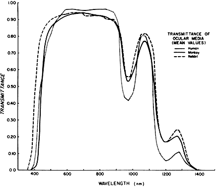
the rhesus monkey, report SAM-TR-78-32. Brook Air Force Base, TX, USAF School of Aerospace Medicine, 1978

53. Weast RC (ed): CRC Handbook of Chemistry and Physics: A Ready Reference Book of Clinical and Physical Data, p 184. Boca Raton, FL, CRC Press, 1984

54. Boettner EA, Wolter JR: Transmission of ocular media. Invest Ophthalmol Vis Sci 1:776, 1962

55. Lerman S: Chemical and physical properties of the normal and aging lens: Spectroscopic (UV, fluorescence, phosphorescence and NMR) analyses. Am J Optom Physiol Optics 64:11, 1987

56. Brancato R, Pratesi R: Application of diode laser in ophthalmology. Laser Ophthalmol 1:119, 1987

57. Azzolini C, Docchio F, Brancato R et al: Interactions between light and vitreous fluid substitutes. Arch Ophthalmol 110:1468, 1992

58. Ham WT, Mueller HA, Ruffolo JJ et al: Solar retinopathy as a function of wavelength: Its significance for protective eyewear. In Williams TP, Baker BN (eds): The Effects of Constant Light on Visual Processes, pp 319–346. New York, Plenum Press, 1980

59. Geeraets WJ, Berry ER: Ocular spectral characteristics as related to hazards from laser and other light sources. Am J Ophthalmol 66:15, 1968

60. Ham WT, Ruffolo JJ, Mueller HA et al: Action spectrum of retinal injury from near UV radiation in the aphakic monkey. Am J Ophthalmol 93:299, 1982

61. Lawhill T: Effects of prolonged exposure of rabbit retina to low intensity light. Invest Ophthalmol Vis Sci 12:45, 1973

62. Flynn HW, Brod RD: Protection from operating microscope-induced retinal phototoxicity during pars plana vitrectomy. Arch Ophthalmol 106:1032, 1988

63. Brod RD, Olson KR, Ball SF et al: The site of operating microscope light induced injury on the human retina. Am J Ophthalmol 207:390, 1989

64. Friedman E, Kuwabara T: Retinal pigment epithelium: Damaging effects of radiant energy. Arch Ophthalmol 80:265, 1968

65. Rinkoff J, Machemer R, Hida T et al: Temperature dependent light damage to the retina. Am J Ophthalmol 102:452, 1986

66. Parver LM, Ham WT, Mitchard et al: Arterial oxygen levels as a risk factor for photochemical retinal damage in the surgical eye patient. Ophthalmology 91(suppl):135, 1984

67. Irvine AR: Retinal complications of cataract surgery. In Freeman WR (ed): Practical Atlas of Retinal Disease and Therapy, pp 155–161. Philadelphia, Lippincott-Raven, 1998

68. Irvine AR, Wook I, Morris AW: Retinal damage from the illumination of the operating microscope: An experiment and study in pseudophakic monkeys. Arch Ophthalmol 102:1358, 1984

69. Colvard DM: Operating microscope light-induced retinal injury: Mechanisms, clinical manifestation and preventive measures. Am Intraocular Implant Soc J 10:438, 1984

70. Griess GA, Blankenstein MF: Additivity and repair of actinic retinal lesions. Invest Ophthalmol Vis Sci 20:803, 1981

71. Roberts JE, Reme CE, Dillon J et al: Exposure to bright light and the concurrent use of photosensitizing drugs. N Engl J Med 326:1500, 1992

72. Davidson PC, Sternberg P Jr: Potential retinal phototoxicity. Am J Ophthalmol 116:497, 1993

73. Cowan CL: Light hazards in the operating room. J Natl Med Assoc 84:425, 1992

74. Borsje RA, Vrensen GF, van Best JA et al: Fluorophotometric assessment of blood-retinal barrier function after white light exposure in the rabbit eye. Exp Eye Res 50:297, 1990

75. Tso MOM, Fine B, Zimmerman L: Photic maculopathy produced by the indirect ophthalmoscope. Am J Ophthalmol 73:686, 1972

76. Tso MOM, Woodward BJ: Effect of photic injury on the retinal tissues. Ophthalmology 90:952, 1983

77. Yannuzzi LA, Fisher YL, Krueger A et al: Solar retinopathy: A photobiological and geophysical analysis. Trans Am Ophthalmol Soc 85:120, 1987

78. Penner R, McNair JN: Eclipse blindness. Am J Ophthalmol 61:1452, 1966

79. Hatfield EM: Eye injuries and the solar eclipse. Sight Saving Rev 40:79, 1970

80. Cordes FC: A type of foveomacular retinitis observed in the US Navy. Am J Ophthalmol 27:803, 1944

81. Ewald RA, Ritchey CL: Sun gazing as the cause of foveomacular retinitis. Am J Ophthalmol 70:491, 1970

82. Kerr LM, Little HL: Foveomacular retinitis. Arch Ophthalmol 76:498, 1966

83. Marlor RL, Blais BR, Preston FR et al: Foveomacular retinitis, an important problem in military medicine: Epidemiology. Invest Ophthalmol Vis Sci 12:5, 1973

84. Wergeland FL Jr, Brenner EH: Solar retinopathy and foveomacular retinitis. Ann Ophthalmol 7:495, 1975

85. Naidoff MA, Sliney DH: Retinal injury from a welding arc. Am J Ophthalmol 77:663, 1974

86. Romanchuk KG, Pollak V, Schneider RJ: Retinal burn from a welding arc. Can J Ophthalmol 13:120, 1978

87. Uniat L, Olk RJ, Hanish SJ: Welding arc maculopathy. Am J Ophthalmol 102:394, 1986

88. Wurdemann HV: The formation of a hole in the macula: Light burn from exposure to electric welding. Am J Ophthalmol 19:457, 1936

89. Henry MM, Henry LM, Henry LM: A possible cause of chronic cystic maculopathy. Ann Ophthalmol 9:455, 1977

90. Macy JI, Baerveldt G: Pseudophakic serous maculopathy. Arch Ophthalmol 101:228, 1983

91. Fishman GA: Light-induced maculopathy from surgical microscopes during cataract surgery. In Ernest JT (ed): 1985 Year Book of Ophthalmology, pp 177–181. Chicago, Year Book Medical Publishers, 1985

92. Byrnes GA, Chang B, Loose I et al: Prospective incidence of photic maculopathy after cataract surgery. Am J Ophthalmol 119:231, 1995

93. Gomolin JES, Koenekoop RK: Presumed photic retinopathy after cataract surgery: An angiographic study. Can J Ophthalmol 28:221, 1993

94. Brod RD, Barron BA, Suelflow JA et al: Phototoxic retinal damage during refractive surgery. Am J Ophthalmol 102:121, 1986

95. Kramer T, Brown R, Lynch M et al: Molteno implants and operating microscope-induced retinal phototoxicity: A clinicopathologic report. Arch Ophthalmol 109:379, 1991

96. Rouland JF, Constantinides G, Turut P: Light-induced maculopathy following epikeratoplasty. Refractive Corneal Surg 6:270, 1990

97. Kelly NE, Wendell RT: Vitreous surgery for idiopathic macular holes: Results of a pilot study. Arch Ophthalmol 109:654, 1991

98. Brod RD, Ball SF, Packer AJ: A model for predicting the site of paraxial lesions secondary to “coaxial” operating microscope illumination system. Am J Ophthalmol 104:516, 1987

99. Postel EA, Pulido JS, Byrnes GA et al: Long-term follow-up of iatrogenic phototoxicity. Arch Ophthalmol 116:753, 1998

100. Hupp SL: Delayed, incomplete recovery of macular function after photic retinal damage associated with extracapsular cataract extraction and posterior lens insertion. Arch Ophthalmol 105:1022, 1987

101. Leonardy NJ, Dabbs CK, Sternberg P Jr: Subretinal neovascularization after operating microscope burn. Am J Ophthalmol 109:224, 1990

102. van der Hoeve J: Eye lesions produced by light rich in ultraviolet rays: Senile cataract, senile degeneration of the macula. Am J Ophthalmol 3:178, 1920

103. Gjessing HGA: Gibt es einen antagonismus zwischen cataracta senilis und haabscher seniler makulaveranderungen? Z Augenheilkd 56:79, 1925

104. Gjessing HGA: Gibt es einen antagonismus zwischen cataracta senilis und haabscher seniler makulaveranderungen? Acta Ophthalmol 31:401, 1953

105. Sperduto RD, Hiller R, Seigel D: Lens opacities and senile maculopathy. Arch Ophthalmol 99:1004, 1981

106. Chumbley LC: Impressions of eye diseases among Rhodesian blacks in Marshonland. S Afr Med J 52:316, 1977

107. Gregor Z, Joffe L: Senile macular changes in the black African. Br J Ophthalmol 62:554, 1978

108. Maltzman BA, Mulvihill MN, Greenbaum A: Senile macular degeneration and risk factors: A case control study. Ann Ophthalmol 11:1197, 1979

109. Hyman LG, Lilienfeld AM, Ferris FL III et al: Senile macular degeneration: A case control study. Am J Epidemiol 118:213, 1983

110. Weiter JJ, Delori FC, Wing GL et al: Relationship of senile macular degeneration to ocular pigmentation. Am J Ophthalmol 99:185, 1985

111. Holz FG, Piquet B, Minassian DC et al: Decreasing stromal iris pigmentation as a risk factor for age-related macular degeneration. Am J Ophthalmol 117:19, 1994

112. Sandberg MA, Gaudio AR, Miller S et al: Iris pigmentation and extent of disease in patients with neovascular age-related macular degeneration. Invest Ophthalmol Vis Sci 35:2734, 1994

113. La Vail MM: Eye pigmentation and constant light damage in the rat retina. In Williams TP, Baker BN (eds): The Effects of Constant Light on Visual Processes, pp 357–387. New York, Plenum, 1980

114. Penn JS, Howard AG, Williams TP: Light damage as a function of “light history” in the albino rat. In La Vail MM, Hollyfield JG, Anderson RE (eds): Retinal Degeneration: Experimental and Clinical Studies, pp 439–447. New York, Alan R Liss, 1985

115. Rapp LM, Williams TP: A parametric study of retinal light damage in albino and pigmented rats. In Williams TP, Baker BN (eds): The Effects of Constant Light on Visual Processes, pp 135–139. New York, Plenum Publishing, 1980

116. Taylor HR, Munoz B, West S et al: Visible light and risk of age-related macular degeneration. Trans Am Ophthalmol Soc 88:163, 1990

117. Taylor HR, Munoz B, West S et al: The long-term effects of visible light on the eye. Arch Ophthalmol 110:99, 1992

118. Cruickshanks KJ, Lein R, Klein BEK: Sunlight and age-related macular degeneration: The Beaver Dam Eye Study. Arch Ophthalmol 111:514, 1993

119. Gottsch JD, Bynoe LA, Harlan JB et al: Light-induced deposits in Bruch's membrane of protoporphyric mice. Arch Ophthalmol 111:126, 1993

120. Gottsch JD, Pou S, Bynoe LA et al: Hematogenous photosensitization: A mechanism for the development of age-related macular degeneration. Invest Ophthalmol Vis Sci 31:1674, 1990

121. Gallard ER, Atherton SJ, Eldred G et al: Photophysical studies on human retinal lipofuscin. Photochem Photobiol 61:448, 1995

122. Harlan JB, Weidenthal DT, Green WR: Histologic study of a shielded macula. Retina 17:232, 1997

123. Drucker BL, Shapiro LA: Protective effect of occlusion on disciform degeneration. Ann Ophthalmol 20:118, 1988

124. Patz A, Hoeck LE, DeLaCruz E: Studies on the effect of high oxygen administration in retrolental fibroplasia: Nursury observations. Am J Ophthalmol 35:1248, 1952

125. Glass P: Light and the developing retina. Doc Ophthalmol 74:195, 1990

126. Glass P, Avery GB, Subramanian KN et al: Effect of bright light in the hospital nursery on the incidence of retinopathy of prematurity. N Engl J Med 313:401, 1985

127. Sadda SR, Yu YS, de Juan E Jr et al: Photosensitization-induced retinopathy in the newborn beagle. Invest Ophthalmol Vis Sci 35:1202, 1994

128. Katz ML, Robinson WG Jr: Autoxidative damage to the retina: Potential role in retinopathy of prematurity. In Flynn JT, Phelps DL (eds): Retinopathy of Prematurity: Problem and Challenge,pp 237–248. New York, Alan R Liss, 1988

129. Riley PA, Slater TF: Pathogenesis of retrolental fibroplasia. Lancet ii:265, 1969

130. Nielson JC, Naash MI, Anderson RE: The regional distribution of vitamins E and C in mature and premature human retinas. Invest Ophthalmol Vis Sci 29:22, 1988

131. Penn JS, Thum LA, Naash MI: Oxygen-induced retinopathy in the rat: Vitamins C and E as potential therapies. Invest Ophthalmol Vis Sci 33:1636, 1992

132. Bynoe LA, Gottsch JD, Sadda SR et al: An elevated hematogenous photosensitizer in the preterm infant. Invest Ophthalmol Vis Sci 34:2878, 1993

133. Robinson J, Mosely MJ, Thompson JR et al: Eyelid opening in preterm neonates. Arch Dis Child 64:943, 1989

134. Robinson J, Fielder AR: Pupillary diameter and reaction to light in preterm neonates. Arch Dis Child 65:35, 1990

135. Isenberg SJ, Molarte A, Vazquez M: The fixed and dilated pupils of premature neonates. Am J Ophthalmol 110:168, 1990

136. Robinson J, Fielder AR: Light and the immature visual system. Eye 6:166, 1992

137. Light Reduction in Retinopathy of Prematurity (LIGHT-ROP) Cooperative Group: Lack of efficacy of light reduction in preventing retinopathy of prematurity. N Engl J Med 338:1620, 1998

138. Calkins JL, Hochheimer BF: Retinal light exposure from operating microscopes. Arch Ophthalmol 97:2363, 1979

139. Jampol LM: Aphakic cystoid macular edema: A hypothesis. Arch Ophthalmol 103:1134, 1985

140. Jampol LM, Kraff MC, Sanders DR et al: Near UV radiation from the operating microscope and pseudophakic cystoid macular edema. Arch Ophthalmol 103:28, 1985

141. Iliff WJ: Aphakic cystoid macular edema and the operating microscope: Is there a connection? Trans Am Ophthalmol Soc 83:476, 1985

142. Kraff MC, Sanders DR, Jampol LM et al: Effect of an ultraviolet filtering intraocular lens on cystoid macular edema. Ophthalmology 92:366, 1985

143. Absolon MJ: The effects of ultraviolet light on the eye. Trans Ophthalmol Soc UK 104:522, 1985

144. Green WR, Robertson DM: Pathologic findings of photic retinopathy in the human eye. Am J Ophthalomol 112:520, 1991

145. Parver LM, Auker CR, Fine BS: Observations on monkey eyes exposed to light from an operating microscope. Ophthalmology 90:964, 1983

146. Jaffe GJ, Irvine AR, Wood IS et al: Retinal phototoxicity from the operating microscope: The role of inspired oxygen. Ophthalmology 95:1130, 1988

147. Gorn RA, Kuwabara T: Retinal damage by visible light. Arch Ophthalmol 77:115, 1967

148. Kuwabara T, Gorn RA: Retinal damage by visible light: An electron microscopic study. Arch Ophthalmol 79:69, 1968

149. Grignolo A, Orzalesi AG, Castellazzo R et al: Retinal damage by visible light in albino rats. Ophthalmologica 157:43, 1969

150. Kuwabara T: Retinal recovery from exposure to light. Am J Ophthalmol 70:187, 1970

151. Tso MOM, LaPiana FG: The human fovea after sun-gazing. Trans Am Acad Ophthalmol Otolaryngol 79:OP 788, 1975

152. Mainster MA: Spectral transmittance of intraocular lenses and retinal damage from intense light sources. Am J Ophthalmol 85:167, 1978

153. Lerman S: Radiant Energy and the Eye. New York, Macmillan, 1980

154. Lerman S: Parker Heath Memorial Lecture: Photosensitizing drugs and their possible role in enhancing ocular toxicity. Ophthalmology 93:304, 1986

155. Clarke CA, Sneddon IB: Nutritional neuropathy in prisoners-of-war and internees from Hong-Kong. Lancet i:734, 1946

156. American National Standard: Requirements for nonprescription sunglasses and fashion eyewear. New York, American National Standards Institute, 1986

157. Borgwardt B, Fishman GA, VanderMeulen D: Spectral transmission characteristics of tinted lenses. Arch Ophthalmol 99:293, 1981

158. Fishman GA: Ocular phototoxicity: Guidelines for selecting sunglasses. Surv Ophthalmol 31:119, 1986

159. Schwartz LK, Norris JL: A corneal shield to prevent light-induced maculopathy during cataract surgery. Am J Ophthalmol 97:658, 1984

160. McIntyre DJ: Phototoxicity: The eclipse filter. Ophthalmology 92:364, 1985

161. Nevyas HJ, Nevyas JY: Surgical corneal light occluder made of black hema. Ophthalmic Surg 16:696, 1985

162. Yanoff M, Kurata F, Lamensdorf M: Inexpensive device to reduce surgical light exposure. Ophthalmology 90(suppl):137, 1983

163. Urinowski E, Cahane M, Ashkenazi I et al: Proximity-sensor dimmer device as an aid in the reduction of operating microscope-induced retinal phototoxicity. Ophthalmic Surg 25:122, 1994

164. Fechner PU, Barth R: Effect on the retina of an air cushion in the anterior chamber and coaxial illumination. Am J Ophthalmol 96:600, 1983

165. Zak R, Jabbour N, Brown S: The effects of retinal hypothermia on argon blue-green laser thresholds in vitrectomized rabbit eyes. Invest Ophthalmol Vis Sci (suppl):292, 1982

166. Ham WT Jr, Mueller HA, Guerry RK: Light damage. In Sheffield JB, Hilfer SR (eds): Cell and Developmental Biology of the Eye: The Microenvironment and Vision, pp 141–158. New York, Springer-Verlag, 1987

Back to Top