Chapter 52
Primary Open-Angle Glaucoma
YOUNG H. KWON and JOSEPH CAPRIOLI
Main Menu   Table Of Contents

Search

DEFINITION
PATHOGENESIS
EPIDEMIOLOGY
DIAGNOSIS
TREATMENT
NEUROPROTECTION: POTENTIAL NEW AVENUES FOR GLAUCOMA TREATMENT
GENERAL MANAGEMENT GUIDELINES
REFERENCES

DEFINITION
Primary open-angle glaucoma (POAG) is the most common form of adult glaucoma. It is an acquired condition characterized by (1) open anterior chamber angle, with normal gonioscopic appearance; (2) chronic progressive loss of retinal ganglion cells, manifest by a characteristic optic neuropathy; and (3) typical patterns of visual field loss in the more advanced stages. The more classic requirement for elevated intraocular pressure (IOP) is usually dropped from the modern definition. Normal (mean ± standard deviation) IOP is 15.5 (±2.57) mmHg.1 Assuming a gaussian distribution in which two standard deviations include the values of about 95% of the population, an IOP of 20.5 mmHg (mean plus 2 standard deviations) could be used as the upper limit of normal IOP. Although the distribution of normal IOP is skewed toward the higher range, this statistical definition was widely accepted and previously used in the definition of POAG.

POAG is an acquired condition, with onset typically after age 40. It affects both eyes but often asymmetrically. In early and even in moderate stages, the patient is usually asymptomatic. Abnormalities of the visual field occur insidiously and initially involve the midperiphery. In more advanced stages, the patient may become aware of an enlarging scotoma, particularly when it encroaches on fixation. If left untreated, vision may be lost. The natural history of the disease is one of a slowly progressive optic neuropathy. It has been estimated that untreated glaucoma can take an average of 14.4 years to progress from early to end stage at IOP of 21 to 25 mmHg, 6.5 years at 25 to 30 mmHg, and 2.9 years at more than 30 mmHg.2

Normal tension glaucoma occurs in a subset of patients who have POAG and can be described as a primary, acquired, progressive optic neuropathy characterized by typical patterns of cupping and visual field changes. It is distinguished from POAG by the absence of IOP elevated above the statistically normal range. There are those who believe that normal-tension glaucoma and POAG do not represent two separate entities but different places in the spectrum of the same disease process. Proponents of the terminology “normal-tension glaucoma” disagree and note some significant clinical differences.3–5

Ocular hypertension refers to IOP elevated above the statistically normal range without evidence of characteristic glaucomatous optic nerve damage or visual field abnormalities. Those with ocular hypertension or optic discs with enlarged cups are often referred to as “glaucoma suspects” because the risk of glaucoma increases with elevated IOP6 or an abnormally large cup (see Epidemiology).

Back to Top
PATHOGENESIS
The pathogenesis of POAG is incompletely understood. It is believed that the elevated IOP in glaucoma results from increased resistance within the aqueous drainage system.7 Histopathologic studies of the outflow structures have uncovered potential mechanisms.8 Other studies have addressed mechanisms of the optic nerve atrophy and cupping in glaucoma.9 The mechanical theory proposes a direct pressure induced-damage to the retinal ganglion cell axons at the level of the lamina cribrosa, whereas the vascular theory proposes microvascular changes and resultant ischemia in the optic nerve head. Cellular and molecular events that could lead to glaucomatous retinal ganglion cell death have also been proposed.10

MECHANISMS OF ELEVATED INTRAOCULAR PRESSURE

Morphologic alterations in the extracellular matrix of the aqueous outflow system in patients with glaucoma have been described in detail.8,11,12 Briefly, these changes include nodular proliferation of extracellular collagen, fragmentation, and “curling” of the collagen fiber bundles. There is an increase in glycosaminoglycan content8 but an overall decrease in hyaluronic acid.13 The endothelial cells lining the trabecular meshwork show “foamy” degeneration with basement membrane thickening.11 Ultrastructural changes in the juxatacanalicular tissue—the outermost aspect of the trabecular meshwork believed to be the most likely site of obstruction in glaucoma—have also been described.8,14–16 There is accumulation of nonfibrillar material with characteristics of basement membrane, curly collagen, and chondroitin sulfate protein complex. Changes in matrix vesicles (extracellular lysosomes), sheath material from subendothelial elastic-like fibers, extracellular glycoprotein, fibronectin, and elastin have been reported.16–20 Specificity of some of the morphologic changes has been questioned because similar findings have been noted in normal, aged eyes without glaucoma.15 This has led some to speculate that glaucomatous changes in the outflow pathway may represent an accelerated aging process.21

In addition to the changes in the trabecular meshwork, collapse of Schlemm's canal has been invoked as another mechanism of outflow obstruction.22,23 To support this hypothesis, adhesions between the inner and outer walls of Schlemm's canal have been shown.8,22 There is a certain amount of segmental variability in histopathologic specimens, however.24

Finally, differences in composition of the aqueous have been suggested as another mechanism for increased outflow resistance. Transforming growth factors (TGF) are polypeptides with multiple cellular regulatory functions. TGF can inhibit epithelial cell proliferation, induce extracellular matrix protein synthesis, and stimulate mesenchymal cell growth. Elevated levels of TGF-β2 have been found in the aqueous of glaucoma eyes.25 The study speculated that increased TGF-β2 levels may be responsible for the decreased cellularity of the trabecular meshwork and may lead to increased debris and resistance to outflow. Others report decreased collagenase activity, increased collagen synthesis, and elevated levels of metalloproteinase-1 inhibitor in the aqueous of glaucoma eyes.26 The study suggests that the decrease in collagen degradation may lead to excess deposition of collagen and loss of the trabecular meshwork cells in glaucoma. Despite these studies, the detailed cellular events and molecular substrates that lead to abnormalities of outflow resistance in glaucoma remain poorly understood.

Molecular genetic studies of large families with juvenile open-angle glaucoma have led to identification of the first glaucoma gene (GLC1A) in chromosome 1.27 Interestingly, about 3% of patients with typical adult-onset POAG also have a mutation in the GLC1A gene.28 This suggests gene mutation is responsible for a small but significant portion of POAG. Cellular and molecular events that lead a defective GLC1A gene and cause elevated IOP and glaucoma remain an active area of research.

MECHANISM OF OPTIC NERVE DAMAGE

Historically, glaucomatous optic nerve damage has been attributed to either a mechanical or vascular etiology. It is unlikely, however, that either theory alone will fully explain the optic nerve damage in glaucoma. POAG likely represents a diverse group of diseases, each involving one or more mechanisms. For the purpose of discussion, these broad mechanisms are considered separately.

MECHANICAL CONSIDERATIONS

Both in vitro and in vivo studies have shown that elevated IOP can cause posterior bowing of the lamina cribrosa, the collagenous structure that supports the retinal ganglion cell axons as they exit the eye.29–31 The lamina cribrosa is made up of about 10 parallel plates, each with various-sized pores that allow bundles of axons to pass through and yet maintain the competence of the eye to hold pressure. Evidence suggests that the plates of the lamina cribrosa are compressed in POAG and may even be entirely collapsed in some cases.32 Such physical distortion of the lamina cribrosa is thought to damage the passing axons by distortion or kinking. Other studies have shown elongation of the pores within the lamina cribrosa, suggesting mechanical forces that may stretch and fragment smaller beams.33 Changes in the extracellular matrix have been described that may lead to the loss of structural support in the lamina cribrosa.34–37 These changes include basement membrane thickening, disorganized and fragmented laminar beams, increased level of certain types of collagen, and structural changes in elastin. Interpretation of these morphologic changes within the lamina cribrosa should be done cautiously because they may represent secondary rather than primary changes in glaucoma.

There is evidence that elevated IOP can impede axoplasmic flow within the retinal ganglion cell axon.38–41 Axonal transport is vital to the normal functioning of neurons; retrograde axonal transport of target-derived neurotrophic factors may be essential for cell survival.42,43 It has been suggested that elevated IOP may lead to the degeneration of retinal ganglion cells by interfering with retrograde axoplasmic flow of essential neurotrophic factors. Lack of neurotrophic factors may trigger apoptosis (programmed cell death) in the retinal ganglion cell (see Cellular Mechanisms of Ganglion Cell Death).

VASCULAR CONSIDERATIONS

Proponents of the vascular theory argue that microvascular changes in the optic nerve head are responsible for glaucomatous optic nerve damage.44 Blood supply to the prelaminar and laminar areas of the optic nerve is derived from the peripapillary choroid and short posterior ciliary arteries.45 The vascular supply to the anterior optic nerve may be compromised in glaucoma by several different mechanisms:

  1. The capillary network of the optic nerve head may be selectively lost in POAG.46 Another
    study, however, showed that the retinal ganglion cell axons and the capillary network are lost at the same rate, suggesting there is no selective loss or pre-existing damage to the capillary network.47
  2. Hayreh noted the importance of the “watershed” zones of the choroidal blood supply.48 The watershed zones refer to the border areas between various choroidal segments, each supplied by a short posterior ciliary artery. The watershed zones represent potential areas of compromised circulation and can include the optic nerve head. In addition, nocturnal systemic hypotension has been proposed as an additional risk factor for the development of glaucoma.49
  3. An epidemiologic association between POAG and systemic microvascular disease (e.g., diabetes mellitus) has been reported.50 Other studies have failed to show a significant correlation between POAG and diabetes, however.51,52
  4. There is some evidence that autoregulation of blood flow in the optic nerve head is altered in POAG.53,54 Autoregulation is an important mechanism by which arterioles dilate or constrict with the rise or fall in perfusion pressure to maintain constant blood flow to the retina. In glaucoma, this autoregulatory mechanism may be defective and may predispose the optic nerve to ischemic damage.

CELLULAR MECHANISMS OF GANGLION CELL DEATH

There is increasing interest in elucidating the cellular and molecular events that lead to retinal ganglion cell death in glaucoma. Apoptosis is a process by which excess neurons undergo spontaneous degeneration during normal development. Apoptosis has been demonstrated in primate55 and rat models of glaucoma.56,57 These studies suggest that elevated IOP may trigger cellular events leading to apoptosis. One hypothesis is that elevated IOP impairs the retrograde axonal transport of essential neurotrophic factors58,59 and in turn triggers apoptosis of the retinal ganglion cell.

Glutamate is an excitotoxic amino acid that normally functions as a neurotransmitter in the retina. Ischemia can produce excess levels of extracellular glutamate, which may lead to cell death through a complex series of cellular events that involves glutamate receptors and Ca+ + influx into the cell.60–62 Elevated levels of glutamate in the vitreous have been demonstrated in glaucomatous monkeys and humans, garnering support for this theory.63 It is unclear whether the accumulation of vitreal glutamate is a primary or secondary process in glaucoma.

Back to Top
EPIDEMIOLOGY
Glaucoma is a significant public health problem. It is the second leading cause of blindness in the U.S.64,65 An estimated 2.25 million Americans have glaucoma and about 150,000 are legally blind.66,67 In the older African-American population, glaucoma is the leading cause of blindness, and the risk of blindness from glaucoma is 6.6 times greater in that population, compared with white Americans.67 In the world, glaucoma is the third leading cause of blindness68; an estimated 13.5 million people may have glaucoma and 5.2 million of those may be blind.68 In Nigeria, up to 34% of glaucoma patients may be blind in both eyes and 91% blind in at least one eye.69

PREVALENCE AND INCIDENCE OF POAG

Population-based studies show that the prevalence of POAG ranges from 0.4% to 8.8% in those older than age of 40 (Table 1). On average, POAG is found in 1.9% of white and 0.58% of Asian populations. In black populations however, the prevalence is significantly higher at 6.7%. Although some of the difference can be attributed to epidemiologic study design and the precise definition of POAG, the significantly higher rates observed in Western African populations probably reflect a fundamental risk factor associated with race (see Risk Factors).


TABLE 1. Population-based Prevalence Studies of Primary Open-Angle Glaucoma


Location (Study, year)AgeRacePrevalence (n = Total Number of Participants)
Ferndale, UK (1966) 7040–75White0.4 (n = 4231)
Framingham, MA, USA (Framingham Eye Study, 1977) 5152–85White3.3 (n = 2675)
Beaver Dam, WI, USA (Beaver Dam Eye Study, 1992) 7143–84White2.1 (n = 4926)
County Roscommon, Ireland (1993) 7250White1.9 (n = 2186)
Rotterdam, The Netherlands (Rotterdam Study, 1994) 7355White1.1 (n = 3062)
Blue Mountains region, Australia (Blue Mountains Eye Study, 1996) 7449White3.0 (n = 3654)
Baltimore, MD, USA (Baltimore Eye Survey, 1991) 6640White1.3 (n = 2913)
Baltimore, MD, USA (Baltimore Eye Survey, 1991) 6640Black4.7 (n = 2395)
St. Lucia, West Indies (1989) 7530–86Black8.8 (n = 1679)
Barbados, West Indies (Barbados Eye Study, 1994) 7640–84Mainly black6.6 (n = 4709)
Japan (1991) 7740Asian0.58 (n = 8126)


The incidence rate of POAG is not precisely known. An annual incidence rate of 0.24% was reported in a Swedish population.78 More recently, population-based studies have addressed this issue (Barbados Eye Study and Baltimore Eye Survey) and are ongoing (1999). Conversely, several studies have reported incidence rates of glaucoma in patients with ocular hypertension (Table 2). Ocular hypertension is a well-known risk factor for the development of glaucoma (see Risk Factors). Reported annual incidence rates vary from 0% to 7%; about 1.7% of ocular hypertensive patients become on average, glaucomatous annually.


TABLE 2. Incidence of POAG in Ocular Hypertension


StudyAverage Annual Incidence Rate (%; n = total number of participants, follow-up period in years)
Armaly (1969) 790.1 (n = 5886, 13)
Norskov (1970) 800 (n = 68, 5)
Perkins (1973) 810.5 (n = 124, 6)
Walker (1974) 821.0 (n = 109, 11)
Wilensky et al (1974) 831.0 (n = 50, 6)
Linnér (1976) 840 (n = 92, 10)
Kitazawa et al (1977) 831.0 (n = 75, 9.5)
David et al (1977) 865.0 (n = 61, 3.3)
Hart et al (1979) 877.0 (n = 92, 5)
Lundberg et al (1987) 881.7 (n = 41, 20)

POAG = primary open-angle glaucoma.


RISK FACTORS

Several risk factors for the development of POAG have been identified based on statistical analysis of population-based prevalence studies. Of these, elevated IOP, older age, black race, and positive family history are most strongly correlated with POAG. Other factors such as myopia, diabetes mellitus, systemic hypertension, and migraine or vasospasm are less strongly associated or their association is not clearly established.

Major Risk Factors

Both clinical and experimental studies suggest a strong correlation between elevated IOP and glaucoma. Clinically, secondary glaucoma with elevated IOP such as chronic angle-closure glaucoma produce optic nerve cupping and atrophy, with visual field loss often indistinguishable from that produced by POAG. Experimentally, elevated IOP in animal models of glaucoma can cause optic nerve cupping and atrophy similar to that seen in POAG.89 Furthermore, population-based studies have demonstrated a strong positive correlation between IOP and POAG (Fig. 1).6 The higher the IOP, the greater the prevalence of POAG.

Fig. 1. Prevalence of primary open angle glaucoma as a function of screening IOP (Caucasian Americans [open circles], African Americans [closed circles]). (Reprinted with permission from Sommer A: Relationship between intraocular pressure and primary open angle glaucoma among white and black Americans. Arch Ophthalmol 109:1090, 1991.)

The Baltimore Eye Survey has also shown a positive correlation between older age and POAG in both white and black Americans (Table 3).66 IOP is a confounding factor, however, because it also increases with age in Western populations.90 It is possible that the higher prevalence of glaucoma seen in older age groups may be due to an increased IOP. The proportion of ocular hypertensive patients who have glaucoma increases with age, however, implying that older age itself is a risk factor (Table 4). How aging predisposes to the development of glaucoma remains unclear but it may contribute to the vulnerability of the optic nerve to damage.


TABLE 3. Prevalence of POAG by Age and Race66


AgeObserved rate/100 in Whites (n = total number screened)Observed rate/100 in Blacks (n = total number screened)
40–490.18 (543)0.95 (632)
50–590.32 (618)3.58 (699)
60–690.77 (915)5.05 (614)
70–792.85 (631)7.74 (349)
801.94 (206)10.89 (101)
Total1.10 (2913)4.18 (2395)

POAG = primary open-angle glaucoma.


TABLE 4. Prevalence of POAG and OcularHypertension by Age 90


AgePOAG (%)Ocula Hypertension (%)Prevalence Ratio of POAG and Ocular Hypertension
<40-2.0-
40–490.022.00.01
50–590.313.50.09
60–690.904.50.20
70–792.805.50.51

POAG = primary open-angle glaucoma.


Black race is another important risk factor (see Tables 1 and 3, Fig. 1). In Baltimore, the prevalence of POAG is 6.6 times higher in African-Americans compared with white Americans.67 Collectively, several population-based studies have also shown that the prevalence of POAG among blacks is consistently higher than among whites (see Table 1). This is true even when glaucoma prevalence is adjusted for IOP (see Fig. 1) and age (see Table 3). The data strongly suggest an inherent predisposition of Western African descent to develop POAG.

Positive family history is another important risk factor. The Baltimore Eye Survey found 3.7 times the risk of developing POAG with positive first-degree relatives, 2.17 times with positive parents, and 1.12 times with positive children.91

MINOR RISK FACTORS

Several clinic-based studies suggest that myopia is more frequent in ocular hypertension and glaucoma than would be expected in a normal population.92–94 The elevated IOP associated with myopia does not fully explain the higher prevalence of glaucoma in myopia, suggesting that myopia itself is a risk factor.95 The mechanisms by which myopia predisposes to the development of POAG remain unclear.

Several studies have reported higher prevalence of diabetes mellitus in POAG.96–100 Other studies such as the Framingham Eye Study and Baltimore Eye Survey, however, failed to find such an association.51,52 The Framingham Eye Study determined diabetes by the presence of diabetic retinopathy, whereas the Baltimore Eye Survey relied on a history of diabetes provided by the patient. Both may have underestimated the true prevalence of diabetes. More recently, the Rotterdam Study reported a significant association between POAG and newly diagnosed diabetes mellitus.50 Although it seems plausible that microvascular changes in diabetes could predispose the optic nerve to the glaucomatous damage, direct experimental evidence for this is still lacking.

Systemic hypertension is another risk factor for POAG.101,102 The Rotterdam study found a significant association between elevated systolic blood pressure and POAG but not with normal-tension glaucoma.50 The Baltimore Eye Survey suggested that this relation may be more complex.103 Although both diastolic and systolic blood pressures are modestly associated, the lower perfusion pressure (blood pressure minus IOP) was most strongly associated with POAG.

The Blue Mountains Eye Study found a weak association between typical migraine and POAG in one age group (age 70 to 79).104 A Japanese study failed to find any association between migraine and POAG.105 In contrast, association between migraine and normal-tension glaucoma has been reported.106 Ischemia from periodic vasospasm leading to glaucomatous optic nerve atrophy remains an attractive hypothesis.

A large optic nerve cup may be a risk factor for the development of POAG. In one study, ocular hypertensive patients who developed visual field defects over a 5-year period had significantly larger cup-to-disc ratios (CDRs), compared with those who did not.107 The concept that a large cup is a risk factor for glaucoma remains somewhat problematic, however, because a large cup can also be a manifestation of glaucomatous damage.

A gene responsible for a hereditary form of juvenile open-angle glaucoma (GLC1A) has been identified.27 Interestingly, about 3% of patients with typical adult-onset POAG also show a mutation in the GLC1A gene.28 As more glaucoma genes are identified, it may be possible in the future to assess the risk of glaucoma based on genetic testing.

PROGNOSTIC FACTORS

There is evidence that IOP is not only a risk factor for glaucoma but also a prognostic factor. Higher IOP is associated with faster progression of glaucoma.2 Indeed, there is evidence to show that lowering IOP slows or halts progression of the disease (see Treatment section).

Black race is another prognostic factor. At the initial time of diagnosis, blacks tend to be younger and have more advanced disease than whites.66,108 Glaucoma progression is more rapid109 and the rate of blindness from glaucoma is higher in blacks than whites.67 It is generally believed that the differences are only partially explained by socioeconomic factors or accessibility to medical care.

Disc hemorrhage is another important prognostic factor.110 In one study of unilateral disc hemorrhage, the eye with the hemorrhage showed greater visual field progression than the fellow eye.111 A new disc hemorrhage in a patient with glaucoma is considered to be a sign of progressive optic nerve damage.

Back to Top
DIAGNOSIS
Clinical diagnosis of POAG is based on history, clinical examination, and visual field testing. Previous ocular and medical histories are important in assessing the risk of having or developing POAG (see Risk Factors). Ocular examination may reveal elevated IOP (more than 21 mmHg). Otherwise, the anterior segment examination is unremarkable with an open normal-appearing anterior chamber angle. The optic nerve shows cupping and there may be associated visual field abnormalities. Other open-angle forms of glaucoma, such as those associated with pseudoexfoliation, pigment dispersion, or trauma should be ruled out.

ELEVATED INTRAOCULAR PRESSURE

The IOP is subject to normal diurnal fluctuation of 3 to 6 mmHg.112 Diurnal variation of more than 10 mmHg is unusual and should raise suspicion for glaucoma.113,114 The most common diurnal pattern is an early morning peak.115 The early morning peak has been correlated with the endogenous adrenocortical steroid level.116 Others have challenged this association by pointing out that IOP decreases during sleep despite absence of similar reduction in the plasma cortisol level.117 The clinical significance of diurnal variation is that it is possible to miss the elevated IOP in patients with POAG with a single, isolated measurement. A diurnal curve or multiple measurements of IOP can be carried out throughout the day to confirm the diagnosis of POAG and explore the possibility of normal pressure glaucoma.

OPTIC DISC AND VISUAL FIELD CHANGES

Normal optic discs show healthy intact neural rims,118,119 with corresponding full visual fields. In the normal disc, the average horizontal CDR is about 0.4 when viewed stereoscopically.120 Estimates of the CDR vary by as much as 0.2 when the same nerve is examined multiple times.121 The CDR in fellow eyes tends to be similar (Fig. 2).122,123 There is a significant correlation of the disc appearance among members of the same family.124,125 Thus, significant asymmetry of the optic disc rim between fellow eyes should increase suspicion of a potentially glaucomatous process (Fig. 3). Estimates of CDR alone do not adequately describe the status of the neural rim, however. Focal thinning of the neural rim is often an early sign of glaucoma. Such focal changes are difficult to describe with CDR estimates. Instead, they are better documented by careful disc drawings and color stereoscopic optic disc photography. Disc drawings should emphasize the status of the neural rim, asymmetry between fellow eyes, and asymmetry between the superior and inferior rims of the same eye. Ultimately, stereoscopic disc photography provides a more objective documentation.

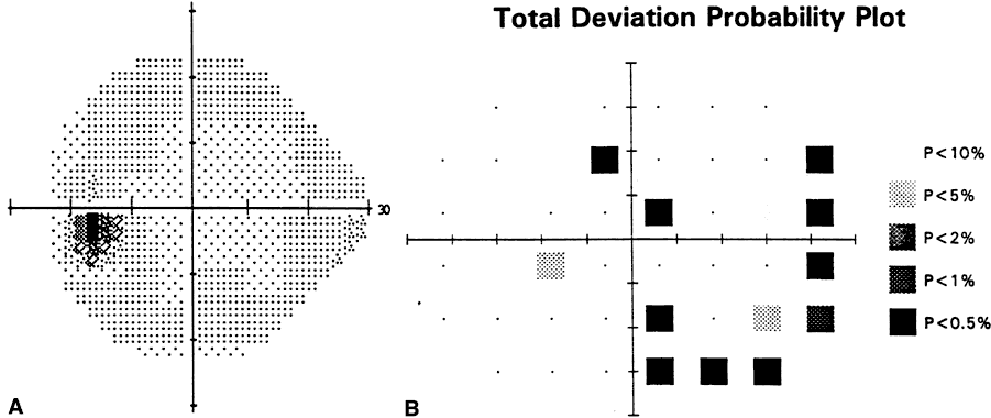
Fig. 2. Normal optic discs and visual fields. A. Right disc B. Left disc of a normal subject. Note the symmetry of cupping and full, intact neural rims. C. Corresponding Humphrey visual field of the right eye and D. Left eye. Both visual fields are within normal limits.

Fig. 3. Asymmetric cupping in early glaucoma. A. Right disc. B. Left disc of a patient with early glaucoma. Note the left disc cup is larger than the right. C. Corresponding Goldmann visual field of the right eye is normal. D. Corresponding visual field of the left eye shows early inferonasal field constriction.

At least four different patterns of glaucomatous optic disc changes have been described.126 They include the focal ischemic, myopic, senile sclerotic, and generalized enlargement of the cup.

The focal ischemic disc shows localized, discrete loss of the neural rim most commonly in the inferotemporal but also in the superotemporal quadrant (Fig. 4). This has also been referred to as “polar notching,” 127 “focal notching,”128 or “pitlike changes.”129 The rest of the neural rim may be relatively intact. The focal changes in the disc have corresponding visual field defects. Dense superior paracentral scotomas are common and are accompanied by pitlike changes in the inferotemporal neural rim. Clinical factors associated with this type of disc are (1) middle- to older age, (2) female gender, (3) normal or elevated IOP, and (4) migraine.126 A third of the focal ischemic type may show disc hemorrhages.

Fig. 4. Focal ischemic disc in glaucoma. A. Note the inferior notching with complete loss of the neural rim and disappearing of the blood vessels into the notch. B. Corresponding Goldmann visual field shows an absolute superior arcuate scotoma.

Myopic discs are tilted discs with temporal crescents and glaucomatous damage to the rim, characterized by superior and inferior thinning of the neural rim (Fig. 5). Their visual field changes are similar to the focal ischemic group but are less likely to threaten fixation or preferentially involve the superior field. These discs tend to be associated with (1) younger age group, (2) male gender, and (3) people of Asian descent.126

Fig. 5. Myopic disc in glaucoma. A. Myopic disc with temporal crescent and inferior notching. B. Corresponding Octopus visual field. Note a dense, superior hemifield defect and early inferonasal step.

Senile sclerotic discs show saucerized and shallow cups with a “moth-eaten” appearance (Fig. 6). They are often accompanied by peripapillary atrophy and choroidal sclerosis, with some pallor of the neural rim. Their visual fields are characterized by relative scotomas with diffuse loss. The senile sclerotic disc is associated with (1) advanced age, (2) normal or elevated IOP, and (3) microvascular diseases such as ischemic heart disease or systemic hypertension.126

Fig. 6. Senile sclerotic disc in glaucoma. A. Saucerized, shallow cup with inferior loss of the neural rim. B. Corresponding Humphrey visual field. Note the diffuse superior field defect, including a paracentral scotoma.

Generalized enlargement of the cup is manifest by diffusely enlarged round cups, without focal loss of the neural rim (Fig. 7). Visual fields show diffuse generalized loss without localized defects. These discs tend to be associated with (1) the younger group, and (2) markedly elevated IOP.126 Disc hemorrhages are infrequently found in this type of disc.

Fig. 7. Generalized enlargement of the cup in glaucoma. A. Generalized enlargement of the cup. B. Corresponding Humphrey visual field. Note the diffuse depression of the threshold sensitivity.

The types of glaucomatous disc changes described generally apply to early and moderate stages of glaucoma. These are examples of relatively “pure” types; many discs have an intermediate appearance. In advanced stages, there is loss of the entire neural rim, and distinctions among different disc types may not be possible (Fig. 8). Histologic cross-section of advanced cupping or “bean-pot” cup shows extreme posterior displacement of the lamina cribrosa and undermining of the disc margin.127,128 The visual field loss in advanced glaucoma may leave only a central or temporal island of remaining vision. In the end, loss of all sight is possible with complete cupping, so-called absolute glaucoma.

Fig. 8. Advanced glaucoma. A. “Bean pod” cupping with almost complete loss of the entire neural rim. B. Corresponding Goldmann visual field shows the loss of central fixation and remaining inferior field.

Splinter hemorrhages at the disc margin are a common feature of glaucoma.130 They occur in all types of glaucomas but are more commonly associated with normal-tension glaucoma.131,324 They frequently occur in the inferior quadrant and are usually seen in the early and moderate stages of glaucoma. They become rare in advanced stages, wherein there is a complete loss of the neural rim.130 Disc hemorrhage is a clinically important sign because its presence has been correlated with an increased rate of optic nerve damage.133 It may also be an early sign of glaucoma because it frequently precedes nerve fiber layer defects,134 focal notches in the neural rim,135 and glaucomatous visual field defects136 (Fig. 9). Loss of retinal ganglion cells and their axons in glaucoma also produces defects in the nerve fiber layer. On ophthalmoscopy, the normal nerve fiber layer appears as fine striations extending temporally in an arcuate fashion from the superior and inferior poles of the disc. With the loss of ganglion cell axons in glaucoma, nerve fiber layer loss can appear as either diffuse or discrete wedge-shaped defects (Fig. 10).137,138 They may follow a disc hemorrhage139 and correlate well with visual field changes.140,141 This finding can be a sensitive and early indicator of glaucoma damage.142–144

Fig. 9. Disc hemorrhage as a sign of glaucoma progression. A. Hemorrhage at the inferotemporal disc margin. B. Corresponding (to A) visual field. C. Thinning of the neural rim and “bayoneting” of a blood vessel at the hemorrhage location 2 years later. D. Corresponding (to C) visual field. Note development of a new superior paracentral scotoma.

Fig. 10. Nerve fiber layer defect in glaucoma. A. Inferior nerve fiber layer wedge defect. B. Corresponding superior visual field defect.

Peripapillary atrophy is also frequently seen in glaucoma (Fig. 11). Peripapillary changes occur more frequently and are more extensive in eyes with glaucoma.145,146 Peripapillary atrophy can also progressively enlarge with progression of glaucoma.147 It is not a specific sign of glaucoma, however, because it is also frequently seen in myopia and aging.

Fig. 11. Peripapillary crescent and atrophy in glaucoma.

DIFFERENTIAL DIAGNOSIS

Glaucoma is not the only condition with optic nerve cupping and visual field loss. Other conditions, both congenital and acquired, can mimic glaucoma. Congenital disc anomalies such as optic nerve coloboma148 (Fig. 12), congenital pit149,150 (Fig. 13), and tilted disc syndrome151 (Fig. 14) can produce optic nerve and visual field changes that are similar to those found in glaucoma. In addition, these anomalies can interfere with recognition of glaucomatous damage when they coexist with glaucoma.151 Acquired conditions such as anterior ischemic optic neuropathy (of the arteritic variety)152,153 and compressive lesions such as intracranial aneurysm154 can produce disc appearance and visual field defects that resemble glaucoma. Young age, highly asymmetric or unilateral disc changes, atypical visual fields, or visual fields that do not correspond to the disc changes should increase the examiner's suspicion of a nonglaucomatous cause.

Fig. 12. Optic disc coloboma and associated visual field deficit can resemble glaucoma. A. Optic disc coloboma. B. Corresponding Humphrey visual field defect. (Courtesy of Wallace L.M. Alward, MD, Iowa City, IA)

Fig. 13. Congenital optic nerve pit and associated visual field deficit can resemble glaucoma. A. Congenital optic nerve pit. B. Corresponding Goldmann visual field defect. (Courtesy of Wallace L.M. Alward, MD, Iowa City, IA)

Fig. 14. Tilted disc can resemble myopic, glaucomatous disc. (Courtesy of Wallace L.M. Alward, MD, Iowa City, IA)

ANCILLARY TESTS

The most widely accepted method for assessment of optic nerve function in glaucoma is perimetry or visual field testing. Both kinetic (e.g., Goldmann perimeter) and static automated perimetry (e.g., Humphrey, Octopus) are standard means for peripheral vision testing. There is evidence that a significant amount of optic nerve damage can occur before standard visual fields become abnormal.155,156 Thus, other functional tests have been developed that can detect glaucoma at earlier stages. These include short-wavelength automated perimetry,157,158 motion detection perimetry (Fig. 15),159 contrast sensitivity,160 and the pattern electroretinogram.161,162

Fig. 15. Early detection of glaucoma using motion perimetry. A. Normal standard Humphrey visual field in a glaucoma suspect. B. Abnormal motion perimetry in the same eye. (Courtesy of Michael Wall, MD, Iowa City, IA)

Sophisticated imaging instrumentation is available to objectively evaluate the status of the optic nerve and nerve fiber layer. It can provide three-dimensional tomographic analysis of the disc based on confocal scanning laser ophthalmoscopy163 and measure the nerve fiber layer thickness with the techniques of scanning laser polarimetry164,165 (Fig. 16) or optical coherence tomography.166 Whether these new instruments will be clinically useful in early detection and accurate monitoring of glaucoma remains to be shown.167

Fig. 16. Correlation between visual field defect and nerve fiber layer thickness. A. Dense, glaucomatous superior arcuate scotoma in the Goldmann visual field. B. Corresponding thinning of the inferior nerve fiber layer thickness as measured by the Nerve Fiber Analyzer GDx (Laser Diagnostic Technologies, Inc., San Diego, CA) in the same eye. Y-axis numbers represent thickness in microns. T, Temporal, S, Superior, N, Nasal, I, Inferior.

Back to Top
TREATMENT
Treatment of POAG is limited to reduction of IOP. This is based on the concept that elevated IOP is at least partly responsible for the optic nerve damage in glaucoma and that IOP reduction can halt or slow the progression of the disease. Numerous clinical studies support this concept.168–173 A recent 5-year prospective study showed a better outcome in eyes treated initially with surgery, compared with eyes treated either with medicine or laser.174 Surgical treatment resulted in lower mean IOP (14.1 mmHg), compared with laser or medical treatment (18.5 mmHg), suggesting that lower IOP helped to protect against disease progression. Additional evidence comes from the Glaucoma Laser Trial and Glaucoma Laser Trail Follow-up Study.174 The lower IOP in the initial laser trabeculoplasty group was associated with less disease progression, compared with the initial medicine group. In 1992, a prospective 4-year clinical study was begun (Early Manifest Glaucoma Trial) to directly address the question of whether IOP reduction, compared with no treatment, makes any difference in newly diagnosed POAG.175

Treatment of POAG involves medications, laser surgery, and incisional surgery.

MEDICAL TREATMENT

Medications used to treat POAG include many classes of drugs, all designed to lower IOP. They include beta-adrenergic antagonists, nonselective adrenergic agonists, selective alpha-2 adrenergic agonists, cholinergic agonists, carbonic anhydrase inhibitors, prostaglandin analogs, and hyperosmotic agents (Table 5).


TABLE 5. Medications Used in the Treatment of Glaucoma


ClassMechanismDrugStrengthDosingCommon Side Effects
Nonselective beta antagonistDecrease aqueous productionTimilol maleate0.25%, 0.5%qd, bidBronchospasm, bradycardia, decrease blood pressure, adversely alter blood lipid profiles, CNS effect (lethargy, confusion, depression, impotence), exacerbate myasthenia gravis, mask hypoglycemic symptoms in diabetics
  Timolol hemihydrate0.25%, 0.5%qd, bid 
  Levobunolol HCL0.25%, 0.5%qd, bid 
  Metipranolol0.3%qd, bid 
  Carteolol HCL1.0%qd, bid 
Selective beta-1 antagonistDecrease aqueous productionBetaxolol HCL0.25%, 0.5%qd, bidLess bronchospasm, but otherwise similar to other beta-blockers
Nonselective adrenergic agonistInitially, decrease aqueous production and increase outflow; later, further increase outflowEpinephrine0.25–2.0%qd, bidSystemic: hypertension, tachycardia, arrhythmia
  Epinephrine HCL1.0–2.0%qd, bidOcular: adrenochrome deposits, drug allergy, follicular conujunctivitis, rebound hyperemia, cystoid macular edemia in aphakia, madarosis
  Epinepryl borate0.5–2.0%qd, bid 
  Epinephrine bitartrate0.5–2.0%qd, bid 
  Dipivefrin HCL0.1%bid 
Alpha-2 agonistDecrease aqueous production, increase uveal outflow (brimonidine)Apraclonidine HCL0.5%, 1.0%bid, tidSystemic: dry mouth, decrease blood pressure, bradycardia
  Brimonidine0.2%bid, tidOcular: follicular conjunctivitis, ocular irritation, blanching, lid retraction, mydriasis, drug allergy (less with brimonidine)
Direct cholinergic agonistIncrease trabecular outflowPilocarpine HCL0.5–6%bid-qidMiosis (decrease vision), brow ache, induced myopia and variable refractive error, exacerbate inflammation, shallow anterior chamber, retinal detachment
  Carbachol0.75-3%bid, tid 
Indirect cholinergic agonist (cholinesterase inhibitor)Increase trabecular outflowEchothiophate iodide0.03–0.25%qd, bidAbove plus, cataractogenic, iris cysts in children, increase pupillary block, prolonged effect of paralyzing agent such as succinylcholine when used concomitantly
  Demecarium iodide0.125%, 0.25%qd, bid 
  Physostigmine0.25–0.5%qd, bid 
  Isofluorophate0.25%qhs 
Carbonic anhydrase inhibitorDecrease aqueous productionDorzolamide (topical)2.0%bid, tidParasthesia of fingers and toes, metallic taste, nausea, malaise,
  Acetazolamide (oral, intravenous)125, 250mgbid, tiddepression, loss of libido, hypokalemia, aplastic anemia, metabolic
   25, 50mgbid, tidacidosis, kidney stones; systemic side effect less with dorzolamide
  Methazolamide (oral)50mgbid, tid 
  Dichlorphenamide (oral)   
Prostaglandin analogueIncrease uveal outflowLatanoprost0.005%qhsIncrease in iris pigment, particularly in hazel iris, cystoid macular edema, hypertrichosis
Hyperosmotic agentDecrease vitreous volumeGlycerine (oral)50, 75%1–1.5 g/kgHeadache, back pain, diuresis, angina, pulmonary edema, heart
  Isosorbide (oral)45%1.5 g/kgfailure, obtundation, seizure, and subarachnoid hemorrhage;
  Mannitol (intravenous)5, 10, 15, 20%1–2 g/kgnausea/vomiting (oral agents)


Topical nonselective beta-adrenergic antagonists such as timolol lower IOP by suppressing aqueous production.176 They inhibit synthesis of cyclic adenosine monophosphate (c-AMP) in the ciliary epithelium and lead to a decrease in aqueous secretion.177 Long-term trials with topical timolol in glaucoma patients have shown a sustained reduction of IOP over time.178,179 Ocular side effects of topical beta-blockers are minor and include burning and decreased corneal sensation. Conversely, systemic side effects can be serious. These include bradycardia; arrhythmia; heart failure; heart block; syncope; bronchospasm or airway obstruction; central nervous system effects (depression, anxiety, weakness, fatigue, or hallucinations); and elevation of blood cholesterol levels.180 A beta-1 selective antagonist, betaxolol, has fewer pulmonary side effects but is also less effective in lowering IOP than the nonselective beta antagonists.181,182 Because of long clinical experience and proved efficacy, topical beta-blockers have assumed a central role in the medical treatment of POAG.

Nonselective adrenergic agonists such as epinephrine lower IOP by several different mechanisms.183 Initially, a vasoconstrictive effect decreases aqueous production; another early effect is an increase in the outflow facility by stimulating c-AMP synthesis. Long-term effects may include a further increase of outflow facility. Clinical efficacy of topical epinephrine appears to be similar184 to or slightly less185 than that of timolol. There are numerous side effects associated with topical adrenergic agonists, including both ocular (burning, reactive hyperemia, adrenochrome deposits, mydriasis, maculopathy in aphakic eyes, corneal endothelial damage, and ocular hypoxia) and systemic (hypertension, tachycardia and arrhythmia) symptoms.186 Dipivefrin is a prodrug that is hydrolyzed to epinephrine as it traverses the cornea.187 It has significantly fewer systemic side effects than epinephrine.188 Generally, the use of adrenergic agonists in the treatment of POAG has declined in recent years with the availability of newer medications that show comparable or better efficacy and have fewer side effects.

Selective topical alpha-2 agonists such as apraclonidine decrease aqueous production.189 In addition, a second generation—the highly selective alpha-2 agonist, brimonidine—appears to increase uveoscleral outflow.190 Clinical studies show that topical apraclonidine and brimonidine are as effective as timolol in reducing IOP (20% to 25%) in patients with glaucoma.191,192 Common ocular side effects include allergic reaction, follicular conjunctivitis, eyelid retraction, mydriasis, and conjunctival blanching.193 Systemically, they can cause headache, dry mouth, fatigue, bradycardia, and hypotension. Long-term use of topical apraclonidine is frequently associated with allergy and tachyphylaxis (decreased effectiveness over time).194 Generally, brimonidine seems to produce fewer ocular side effects than apraclonidine.192

Topical cholinergic agonists such as pilocarpine increase the trabecular outflow by contraction of the longitudinal ciliary muscle.195 The same action may also decrease uveoscleral outflow through the ciliary muscle.196 There are two types of cholinergic agonists: direct and indirect. The direct agents (e.g., pilocarpine) are cholinergic receptor agonists; the indirect agents (e.g., echothiophate iodide) inhibit cholinesterase and prolong the action of native acetylcholine. Clinical efficacy of pilocarpine is comparable to timolol,197 whereas that of echothiophate iodide may be superior to timolol in aphakic patients.198 Systemic side effects of pilocarpine are rare. In contrast, ocular side effects are common and include brow ache, induced myopia, miosis (leading to decreased vision), shallowing of the anterior chamber, retinal detachment, corneal endothelial toxicity, breakdown of the blood-brain barrier (which can exacerbate inflammation), hypersensitivity or toxic reaction, cicatricial pemphigoid of the conjunctiva, and atypical band keratopathy. The indirect agents have ocular side effects that are generally more intense than those of the direct agents. In addition, indirect agents can cause iris cysts in children and cataract in adults. Finally, prolonged respiratory paralysis may occur during general anesthesia in patients who are on cholinesterase inhibitors because of their inability to metabolize paralytic agents such as succinylcholine.199 Use of cholinergic agents (as with nonselective adrenergic agonists) has declined in recent years with the availability of newer medications that have comparable efficacy and fewer side effects.

In the ciliary processes, formation of bicarbonate is linked to Na+ secretion and aqueous humor production. Carbonic anhydrase inhibitors such as acetazolamide reduce aqueous production by decreasing bicarbonate production.200 Another proposed mechanism is related to the metabolic acidosis produced by carbonic anhydrase inhibitors that can reduce aqueous production.201 Acetazolamide, a systemic carbonic anhydrase inhibitor, can decrease aqueous flow by 27% in human eyes.202 Many side effects are associated with systemic carbonic anhydrase inhibitors, including transient myopia; parasthesia of the fingers, toes, and perioral area; urinary frequency; metabolic acidosis; malaise; fatigue; weight loss; depression; potassium depletion; gastrointestinal symptoms; renal calculi formation; and rarely, blood dyscrasia.203 Dorzolamide, a topical carbonic anhydrase inhibitor, has recently become available. It has significantly fewer systemic side effects than oral carbonic anhydrase inhibitors and still has clinical efficacy comparable to that of timolol.204

A prostaglandin analogue, latanoprost, represents the newest class of medications. Latanoprost increases uveoscleral outflow by changing the structure of the ciliary muscle.205 In clinical trials, topical latanoprost was more effective in IOP reduction (by 35%) than timolol (27% reduction).206 Ocular and systemic side effects were well-tolerated except for a 12% incidence of increased iris pigmentation. This curious side effect tends to occur in eyes with mixed green-brown or blue-brown irides.207 The clinical significance of this remains unclear; further studies are needed to understand the cellular nature of this side effect. Use of latanoprost has also been associated with anterior uveitis and cystoid macular edema in susceptible individuals.208

Finally, hyperosmotic agents such as oral isosorbide can rapidly lower IOP by decreasing vitreous volume. They do not cross the blood-ocular barrier and therefore exert oncotic pressure that dehydrates the vitreous. Both oral and intravenous agents are available. Side effects associated with the hyperosmotic agents can be severe and include headache, back pain, diuresis, circulatory overload with angina, pulmonary edema and heart failure, and central nervous system effects such as obtundation, seizure, and cerebral hemorrhage.209 Because of the frequent and potentially serious side effects, they are not used as a long-term agent. They are often used to temporarily reduce high IOP until more definitive treatments can be rendered.

SURGICAL TREATMENT

Surgical treatment of POAG includes laser trabeculoplasty, filtering procedures (full-thickness and guarded procedures), aqueous drainage implants, and cyclodestructive procedures.

LASER TRABECULOPLASTY

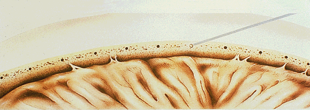
Argon laser application to the trabecular meshwork (argon laser trabeculoplasty) has been shown to significantly lower IOP (Fig. 17).210 The mechanism by which laser trabeculoplasty lowers IOP is not completely understood. Collagen shrinkage and scarring of the trabecular meshwork may open adjacent intertrabecular spaces and decrease the overall outflow resistance.211 Other studies suggest that an increase in mitosis and phagocytosis of the trabecular endothelial cells after laser trabeculoplasty may enhance outflow.212 Argon laser trabeculoplasty can lower IOP in 50% of eyes at 5 years with an attrition rate of 6% to 10% per year.213,214 Prognostic factors for favorable outcome include preoperative IOP of 20 to 29 mmHg, phakic eyes, and older age (older than 40).197,198

Fig. 17. Argon laser trabeculoplasty. (Reprinted with permission from Van Buskirk EM: Clinical Atlas of Glaucoma. Fig. 47-1. Philadelphia, WB Saunders, 1986)

Complications of argon laser trabeculoplasty include discomfort, acutely elevated IOP, progressive visual field loss, peripheral anterior synechiae, iritis, sector palsy of the pupillary sphincter, corneal abrasion, corneal edema, endothelial damage, and vasovagal reaction.217 Transiently elevated IOP of less than 10 mmHg can occur in up to 50% of those treated with laser trabeculoplasty.218 Preoperative treatment with apraclonidine219 or brimonidine220 can significantly reduce the rate of postoperative IOP elevation. Rarely, persistently elevated IOP after laser trabeculoplasty may require trabeculectomy.

FILTERING SURGERY

Full-thickness filtering procedures were designed to create a direct fistula between the anterior chamber and the subconjunctival space, thus bypassing the eye's outflow structures. This can be achieved by thermal cautery,221,222 scleral punch (posterior lip sclerectomy),223 or external trephination.224 Full-thickness filtering procedures effectively lower IOP but are associated with significant postoperative complications, including flat anterior chamber, hypotony, choroidal detachment, endophthalmitis, and cataract formation.

In 1968, a successful partial-thickness guarded-filtering procedure (or trabeculectomy) was first reported.225 Since then, trabeculectomy has become the surgical procedure of choice because of the significantly lower incidence of postoperative complications and comparable efficacy. Clinical success of trabeculectomy approaches 85% to 95% at 2 years.226 Introduction of antifibrotic agents such as 5-fluorouracil and mitomycin C have improved the outcome of trabeculectomy. Antifibrotic agents inhibit wound healing and promote the patency of the fistula (Fig. 18). Trabeculectomy with adjunctive 5-fluorouracil results in significantly lower IOP, compared with trabeculectomy without 5-fluorouracil.227 Adjunctive use of mitomycin C in trabeculectomy produces a similar rate of success as 5-fluorouracil.228 Complications of trabeculectomy are similar to those of full-thickness procedures; they include decreased vision, choroidal effusion, shallow or flat anterior chamber, persistent inflammation, filtration failure, corneal dellen, suprachoroidal hemorrhage, endophthalmitis, chronic hypotony, and maculopathy. Generally, however, trabeculectomy is associated with fewer complications than full-thickness procedures. The risk of persistent hypotony and associated complications such as maculopathy, late-onset bleb leaks, infection of the bleb, and endophthalmitis are increased with the adjunctive use of antifibrotic agents.229,230 The risk of hypotony maculopathy is particularly high in young myopic patients with low scleral rigidity.231 Corneal and conjunctival epithelial toxicity are associated with repeated subconjunctival injections of 5-fluorouracil.232

Fig. 18. Filtering bleb after mitomycin C-augmented trabeculectomy. Mitomycin C-treated bleb is often avascular, thin, and elevated. (Courtesy of Wallace L.M. Alward, MD, Iowa City, IA)

AQUEOUS DRAINAGE IMPLANTS

Use of aqueous drainage implants (or setons) are generally reserved for patients who have complicated secondary glaucomas such as uveitic glaucoma, neovascular glaucoma, or glaucoma after other ocular procedures. In POAG, the drainage devices are typically used when previous trabeculectomy has failed or in an aphakic or pseudophakic eye in which the conjunctiva is excessively scarred for successful trabeculectomy. Broadly, there are two types of drainage implants: nonrestrictive and restrictive (Fig. 19). Molteno and Baerveldt setons are nonrestrictive drainage implants that permit free flow of aqueous from the anterior chamber to a scleral plate through silicone tubing. Restrictive drainage implants such as the Krupin and Ahmed devices incorporate a valvular mechanism that provides some resistance to aqueous outflow and can reduce the incidence of early postoperative hypotony and shallow anterior chamber.

Fig. 19. Glaucoma drainage implant devices. From left to right: Ahmed glaucoma valve (New World Medical, Inc., Rancho Cucamonga, CA); 200-mm2 Baerveldt implant (Iovision, Irvine, CA); Krupin eye disk (Hood Laboratories, Pembroke, MA); Molteno implant (IOP, Inc., Costa Mesa, CA); and OptiMed pressure regulator (OptiMed, Inc., Santa Barbara, CA). (Reprinted with permission from Prata JA, Mérmoud A, LaBree L, Minckler DS: In vitro and in vivo flow characteristics of glaucoma drainage implants. Ophthalmology 102:894, 1995; Courtesy of Ophthalmology)

Surgical outcome of the Molteno seton has a success rate of 60% to 80% at 6 months.233 There is evidence that the double-plate Molteno implant with larger surface area lowers IOP more than the single-plate implant.234 The Baerveldt seton produces surgical results similar to the Molteno.235 Both the Krupin and Ahmed valves achieved similar surgical results at 1 year.236,237 Complications of the aqueous drainage implants include hypotony, choroidal detachment and flat anterior chamber.238 Temporary reduction of flow with the application of a dissolvable suture ligature around the tube until scleral plate encapsulation occurs can reduce complications related to early postoperative hypotony. Early postoperative hypotony and shallow anterior chamber seem less frequently associated with restrictive devices such as the Ahmed,237 although long-term IOP control may be better with the large-plate nonrestrictive devices. Other complications include inflammation, tube obstruction, elevated IOP, tube migration, implant and tube erosion, corneal decompensation, cataract, endophthalmitis, strabismus, and epithelial downgrowth.

CYCLODESTRUCTIVE PROCEDURES

If the patient is a poor candidate for trabeculectomy or drainage implant surgery, a cyclodestructive procedure can be considered. Cyclodestructive procedures decrease aqueous production by ablating the portion of the ciliary body that produces aqueous. This can be achieved by freezing the ciliary epithelium and capillaries within the ciliary body (cyclocryotherapy).239,240 Cyclocryotherapy was shown to be effective in controlling IOP in aphakic glaucoma and glaucoma after penetrating keratoplasty.241 Laser has also been used to selectively destroy the ciliary processes (cyclophotocoagulation). Two commonly used lasers for this purpose are transscleral Nd:YAG and semiconductor-diode lasers. These lasers can produce thermal damage to the ciliary processes and decrease aqueous production. Nd:YAG cyclophotocoagulation has been shown to reduce IOP by 44% to 68%.242–244 Diode cyclophotocoagulation can achieve IOP control in 60% to 80% of eyes treated (Fig. 20).245 Finally, transpupillary cyclophotocoagulation246 and more recently, intraocular cyclophotocoagulation with endoscopic visualization, have also been described.247 Complications of cyclodestructive procedures include pain, reduction of visual acuity, inflammation, transient IOP rise, hyphema, vitreous hemorrhage, cataract, hypotony, choroidal detachment, flat anterior chamber, and phthisis.248,249 Generally, cyclophotocoagulation seems to be associated with better tolerance and fewer complications than cyclocryotherapy.

Fig. 20. Contact diode laser transscleral cyclophotocoagulation using the Iris Medical Instruments G-probe (Iris Medical Instruments, Mountain View, CA. Courtesy of Wallace L.M. Alward, MD, Iowa City, IA)

Back to Top
NEUROPROTECTION: POTENTIAL NEW AVENUES FOR GLAUCOMA TREATMENT
The search for neuroprotective agents for glaucoma treatment is grounded in desperation: the desperation of continuing visual loss in some patients despite IOP reduction to quite low levels. Some cases continue to progress despite dramatic IOP lowering to 5 to 10 mmHg. We present possible avenues for neuroprotection, given the likely players in the damage process. This topic is the subject of a recent review.61

CALCIUM CHANNEL BLOCKADE

Calcium channel blockers have been used empirically to treat low-tension glaucoma. Patients with vasospastic conditions and normal pressure glaucoma have been particularly targeted.250,251 Nifedipine and nimodipine have both been used for treatment of normal-tension glaucoma. Blockade of calcium channels at the neuronal cellular level—by interrupting the cascade of events that lead to death from ischemia—is also a reasonable rationale. The systemic lowering of blood pressure by calcium channel blockers is a concern because it could reduce perfusion pressure to the anterior optic nerve head and lead to ischemia. A retrospective clinical study compared 56 patients with glaucoma who were concurrently taking calcium channel blockers to a control group not taking such medications for a mean follow-up period of 3.4 years, suggested that calcium channel blockers may be useful in the management of low-tension glaucoma.252

GLUTAMATE BLOCKADE

Neuronal injury from glutamate receptor-mediated neurotoxicity has been implicated as a central mechanism in a wide variety of central nervous system diseases, including ischemia, trauma, and some chronic neurodegenerative diseases. Excitotoxicity may also interact with other pathophysiologic processes to enhance or facilitate neuronal damage. The possibility that excitotoxicity may play a role in the chronic neurodegeneration of glaucomatous damage has been suggested. Recent reports of elevated levels of glutamate in the vitreous of glaucomatous monkeys and humans have provided additional fuel for this hypothesis.253,254 Whether the high vitreous levels of glutamate are a cause or result of damage is undetermined but high concentrations of this neurotoxin is toxic to the inner retina.

NEUROTROPHINS

The viability of retinal ganglion cells depends on the retrograde flow of neurotrophic compounds from the target tissue to the cell body. Interruption of retrograde axonal transport could interrupt this supply and trigger apoptotic cell death. This concept has recently been reviewed.10 Restoration of these neurotrophic factors by exogenous administration—or more likely, through forms of gene therapy—could restore “sick” retinal ganglion cells back to health.

HEAT-SHOCK PROTEINS

Recent advances in the understanding of the biochemical and molecular biologic events that lead to neuronal cell death have suggested novel therapeutic approaches. Relatively little attention has been drawn to the importance of intrinsic neuroprotective events in the modulation of cell injury. In this context, heat-shock proteins are likely to play an important role in cell survival after a variety of metabolic insults. A recent study showed that retinal ganglion cells express the 72-kd heat-shock protein after hyperthermia, sublethal hypoxia, and glutamate exposure in vitro.255 Furthermore, retinal ganglion cells in culture treated with hyperthermia or sublethal hypoxia were less to susceptible to subsequent damage from excitotoxicity and anoxia. The neuroprotective effect of the induction of heat-shock protein synthesis by hyperthermia and sublethal hypoxia suggests a role for heat-shock protein as a protective mechanism against ischemic and excitotoxic retinal ganglion cell death.

NITRIC OXIDE SYNTHASE INHIBITION

Nitric oxide is a rapidly diffusing gas with a short half-life in vivo. It has a vasodilatory action and may act as a nonconventional neurotransmitter in the brain. Nitric oxide in sufficient concentrations is a potent neurotoxin. The exact place of nitric oxide in the cascade of events associated with ischemic central nervous system damage is not known but it likely plays an important role. Inhibitors of nitric oxide synthase can protect neurons from nitric oxide toxicity.256

ANTIOXIDANTS

The reperfusion phase after ischemic injury produces highly reactive compounds called free radicals. These oxygen-containing molecules have unpaired electrons and react with lipids, nucleic acids, and proteins. They are thought to be important mediators of reperfusion injury. Free radicals may also facilitate the release of excitotoxins, and both may work together to bring about cellular death from ischemia.257 Free radicals recently have been implicated in the slow chronic neurodegeneration of amyotrophic lateral sclerosis,258 so their role in a chronic neural degeneration like glaucoma is entirely feasible. Free-radical scavengers include endogenous enzymes such as catalase and superoxide dismutase and the antioxidant vitamins, especially C and E. Therapy could take the form of turning on the synthesis of endogenous compounds or providing exogenous ones. Some level of antioxidation can be achieved through vitamin therapy but requires well-controlled clinical studies to determine efficacy.

ANTI-APOPTOSIS

Apoptosis is a term applied to suicidal cell death. This is a programmed gene-directed self-destruction that is a normal occurrence in neural development and differentiation. In the adult nervous system of mammals, it is a mechanism of pathologic cell death. Apoptosis appears to occur—at least to some degree—in most models of neural cell death, whether it is caused by axotomy, ischemia, excitotoxicity, or deprivation of neurotrophins. Apoptosis has been shown to be at least one of the mechanisms for retinal ganglion cell death in animal models of pressure-induced glaucoma in the monkey55 and the rat.57 Evidence of necrotic cell death has not been found in human glaucoma or in the monkey model and is the basis for the hypothesis that apoptosis is the dominant mechanism of retinal ganglion cell death in glaucoma. Because the rate of glaucomatous damage is so slow, however, few cells are expected to be found in the agonal phase.

Drugs are being developed to block apoptosis. Deprenyl, originally developed as a monoamine oxidase inhibitor, increases the gene expression that inhibits apoptosis. Other drugs inhibit the later steps of apoptosis, which include the action of proteases on cell proteins. Several antiapoptosis agents have already been evaluated in the rat retina to prevent light-induced death of photoreceptor cells. Intravitreal injections of flunarizine and aurintricarboxylic acid259 can delay apoptotic death of photoreceptors. Much remains to be learned about the mechanisms that initiate and regulate the process of apoptosis in adult mammals.

OPTIC NERVE REGENERATION

Impressive results reported by Aguayo and coworkers demonstrate the feasibility of central nervous system regeneration. Implantation of peripheral nerve sheath grafts into the eyes of rats promotes the regrowth of axotomized retinal ganglion cells into the graft.260 These regenerated axons also have the ability to establish synaptic connections at target cells.261 The peripheral nerve sheath appears to confer on the central neurons the ability to regenerate by providing a suitable environment and growth factors. This approach may yield important molecular insights into neuroprotection or neuroregeneration, although it is unlikely to yield any clinically applicable therapies in the near future.

Back to Top
GENERAL MANAGEMENT GUIDELINES
Once the diagnosis of POAG is made, it is usually helpful to set a target IOP. The target IOP is the level at which no further glaucoma progression is expected. Generally, the more advanced the glaucoma damage, the lower the target IOP should be. Therefore, the target IOP is not an absolute value. It varies among patients and even in a specific patient, depending on the previous ocular history, age, status of the optic nerve, associated risk factors, and response to treatment. For example, in mild glaucoma with minimal optic nerve changes and a normal visual field, the target IOP may be 21 mmHg; in advanced glaucoma with marked disc cupping and severe visual field loss, it may be 12 mmHg or even lower.

Once the target IOP is set, treatment to lower IOP is rendered. Glaucoma medications are often prescribed first and may be a beta-blocker, prostaglandin analog, alpha-2 agonist, or topical carbonic anhydrase inhibitor. Other medications such as cholinergic agonists, nonselective adrenergic agonists, or oral carbonic anhydrase inhibitors may also be chosen. Selection of a particular medication depends on efficacy, side effects, patient tolerance, cost, and compliance. Periodic regular follow-up is needed to evaluate these factors. Ineffective medications should be stopped and if necessary, additional medications added until the target pressure is reached. Laser trabeculoplasty can be considered when multiple medications have failed to control IOP. Trabeculectomy can be considered if the laser trabeculoplasty or medications failed to control IOP. Adjunctive antifibrotic agents may be used if the trabeculectomy has a high risk of failure or if a low IOP is desired. If the patient is not a candidate for trabeculectomy, an aqueous drainage implant can be considered. Finally, a cyclodestructive procedure may be necessary if other forms of therapy fail or visual prognosis is poor.

The particular sequence of treatment modalities outlined above represent the authors' bias. Others have suggested that initial treatment with laser174,262 or surgery169,263 may be more effective than medications. Most practitioners, however, still choose at least a trial of medications over laser or surgery as initial therapy in most patients.264,265

The glaucoma patient requires regular follow-up examinations, from 2 to 4 times a year or more often, as indicated by the clinical picture. Formal visual field testing is repeated every 4 to 12 months, depending on the stability of the disease. If unstable or deteriorating, the target IOP may need to be lowered and more aggressive treatment applied.

It is important that the physician treating glaucoma keep the whole patient in mind. Treatment for glaucoma, whether medical or surgical, can significantly impact the patient's quality of life. Treating glaucoma at the expense of the patient's overall health may be counterproductive. Therefore, treatment of glaucoma should be individualized according to the needs and desires of a properly informed patient.

Back to Top
REFERENCES

1. Leydhecker W, Akiyama K, Neumann HG: Der intraokulare Druck gesunder menschlicher Augen. Klin Monatsbl Augenheilkd 133:662, 1958

2. Jay JL, Murdoch JR: The rate of visual field loss in untreated primary open angle glaucoma. Br J Ophthalmol 77:176, 1993

3. Caprioli J, Spaeth GL: Comparison of visual field defects in the low-tension glaucomas with those in the high-tension glaucomas. Am J Ophthalmol 97:730, 1984

4. Chauhan B, Drance SM: The influence of intraocular pressure on visual field damage in patients with normal tension and high tension glaucoma. Arch Ophthalmol 103:1145, 1990

5. Kass MA: Normal-pressure glaucoma. Am J Ophthalmol 125:242, 1998

6. Sommer AE, Tielsch JM, Katz J et al: Relationship between intraocular pressure and primary open angle glaucoma among white and black Americans. Arch Ophthalmol 109: 1090, 1991

7. Grant WM: Further studies on facility of flow through the trabecular meshwork. Arch Ophthalmol 60:523, 1958

8. Li Y, Yi Y: Histochemical and electron microscopic studies of the trabecular meshwork in primary open-angle glaucoma. Eye Sci 1:17, 1985

9. Fechtner RD, Weinreb RN: Mechanisms of optic nerve damage in primary open angle glaucoma. Surv Ophthalmol 39:23, 1994

10. Nickells RW: Retinal ganglion cell death in glaucoma: The how, the why and the maybe. J Glaucoma 5:345, 1996

11. Ashton N: The exit pathway of the aqueous. Trans Ophthalmol Soc UK 80:397, 1960

12. Speakman JS, Leeson TS: Site of obstruction to aqueous outflow in chronic simple glaucoma. Br J Ophthalmol 46:321, 1962

13. Knepper PA, Covici S, Fadel JR et al: Surface-tension properties of hyaluronic acid. J Glaucoma 4:194, 1995

14. Rodrigues MM, Spaeth GL, Sivalingam E et al: Value of trabeculectomy specimens in glaucoma. Ophthalmic Surg 9(2):29, 1978

15. Segawa K: Electron microscopic studies of the trabecular meshwork in primary open-angle glaucoma. Ann Ophthalmol 11:49, 1979

16. Alvarado JA, Yun AJ, Murphy CG: Juxtacanalicular tissue in primary open angle glaucoma and in nonglaucomatous normals. Arch Ophthalmol 104:1517, 1986

17. Rohen JW: Presence of matrix vesicles in the trabecular meshwork of glaucomatous eyes. Graefes Arch Clin Exp Ophthalmol 218:171, 1982

18. Rohen JW: Why is intraocular pressure elevated in chronic simple glaucoma? Ophthalmology 90:758, 1983

19. Babizhayev MA, Brodskaya MW: Fibronectin detection in drainage outflow system of human eyes in ageing and progression of open-angle glaucoma. Mech Ageing Dev 47:145, 1989

20. Umihira J, Nagata S, Nohara M et al: Localization of elastin in the normal and glaucomatous human trabecular meshwork. Invest Ophthalmol Vis Sci 35:486, 1994

21. Fine BS, Yanoff M, Stone RA: A clinicopathologic study of four cases of primary open-angle glaucoma compared to normal eyes. Am J Ophthalmol 91:88, 1981

22. Nesterov AP, Batmanov YE: Trabecular wall of Schlemm's canal in the early stage of primary open-angle glaucoma. Am J Ophthalmol 78:639, 1974

23. Moses RA, Grodski WJ Jr, Etheridge EL et al: Schlemm's canal: the effect of intraocular pressure. Invest Ophthalmol Vis Sci 20:61, 1981

24. Buller C, Johnson D: Segmental variability of the trabecular meshwork in normal and glaucomatous eyes. Invest Ophthalmol Vis Sci 35:3841, 1994

25. Tripathi RC, Li J, Chan WFA et al: Aqueous humor in glaucomatous eyes contains an increased level of TGF-β2. Exp Eye Res 59:723, 1994

26. González-Avila G, Ginebra M, Hayakawa T et al: Collagen metabolism in human aqueous humor from primary open-angle glaucoma. Decreased degradation and increased biosynthesis play a role in its pathogenesis. Arch Ophthalmol 113:1319, 1995

27. Stone EM, Fingert JH, Alward WLM et al: Identification of a gene that causes primary open angle glaucoma. Science 275:668, 1997

28. Alward WLM, Fingert JH, Johnson AT et al: The phenotype of primary open angle glaucoma patients with mutations in the GLC1A gene. Invest Ophthalmol Vis Sci Abstr 38:4335, 1997

29. Levy NS, Crapps EE, Bonney RC: Displacement of the optic nerve head. Response to acute intraocular pressure in primate eyes. Arch Ophthalmol 99:2166, 1981

30. Zeimer RC, Ogura Y: The relation between glaucomatous damage and optic nerve head mechanical compliance. Arch Ophthalmol 107:1232, 1989

31. Coleman AL, Quigley HA, Vitale S et al: Displacement of the optic nerve head by acute changes in intraocular pressure in monkey eyes. Ophthalmology 98:35, 1991

32. Quigley HA, Hohman RM, Addicks EM et al: Morphologic changes in the lamina cribrosa correlated with neural loss in open-angle glaucoma. Am J Ophthalmol 95:673, 1983

33. Miller KN, Quigley HA: The clinical appearance of the lamina cribrosa as a function of the extent of glaucomatous optic nerve damage. Ophthalmology 95:135, 1988

34. Morrison JC, Dorman-Peace ME, Dunkelberger GR et al: Optic nerve head extracellular matrix in primary optic atrophy and experimental glaucoma. Arch Ophthalmol 108:1020, 1990

35. Hernandez MR, Andrzejewska WM, Neufeld AH: Changes in the extracellular matrix of the human optic nerve head in primary open-angle glaucoma. Am J Ophthalmol 109: 180, 1990

36. Hernandez MR: Ultrastructural immunocytochemical analysis of elastin in the human lamina cribrosa. Changes in elastic fibers in primary open-angle glaucoma. Invest Ophthalmol Vis Sci 33:2891, 1992

37. Quigley HA, Brown A, Dorman-Pease ME: Alterations in elastin of the optic nerve head in human and experimental glaucoma. Br J Ophthalmol 75:552, 1991

38. Minckler DS, Bunt AH, Johanson GW: Orthograde and retrograde axoplasmic transport during acute ocular hypertension in the monkey. Invest Ophthalmol Vis Sci 16: 426, 1977

39. Minckler DS, Bunt AH, Klock IB: Radioautographic and cytochemical ultrastructural studies of axoplasmic transport in the monkey optic nerve head. Invest Ophthalmol Vis Sci 17:33, 1978

40. Quigley HA, Anderson DR: The dynamics and location of axonal transport blockade by acute intraocular pressure elevation in primate optic nerve. Invest Ophthalmol 15: 606, 1976

41. Radius RL, Bade B: Pressure-induced optic nerve axonal transport interruption in cat eyes. Arch Ophthalmol 99: 2163, 1981

42. Pearson HE, Stoffler DJ: Retinal ganglion cell degeneration following loss of postsynaptic target neurons in the dorsal lateral geniculate nucleus of the adult cat. Exp Neurol 116:163, 1992

43. Schultz M, Raju T, Ralston G et al: A retinal ganglion cell neurotrophic factor purified from the superior colliculus. J Neurochem 55:832, 1990

44. Hayreh SS: Pathogenesis of optic nerve head changes in glaucoma. Semin Ophthalmol 1:1, 1986

45. Hayreh SS: Structure and blood supply of the optic nerve. In Heilmann K, Richardson KT (eds): Glaucoma: Conceptions of a Disease, p 78. Stuttgart, Thieme, 1978

46. François J, Neetens A: Vascularity of the eye and the optic nerve in glaucoma. Arch Ophthalmol 71:219, 1964

47. Quigley HA, Hohman RM, Addicks EM et al: Blood vessels of the glaucomatous optic disc in experimental primate and human eyes. Invest Ophthalmol Vis Sci 25:918, 1984

48. Hayreh SS: Progress in the understanding of the vascular etiology of glaucoma. Curr Opin Ophthalmol 5:11, 1994

49. Hayreh SS, Zimmerman MB, Podhajsky P et al: Nocturnal arterial hypotension and its role in optic nerve head and ocular ischemic disorders. Am J Ophthal 117:603, 1994

50. Dielemans I, de Jong PTVM, Stolk R et al: Primary open-angle glaucoma, intraocular pressure, and diabetes mellitus in the general elderly population. The Rotterdam Study. Ophthalmology 103:1271, 1996

51. Kahn HA, Leibowitz HM, Ganley JP et al: The Framingham Eye Study. I. Outline and major prevalence findings. Am J Epidemiol 106:17, 1977

52. Tielsch JM, Katz J, Quigley HA et al: Diabetes, intraocular pressure, and primary open-angle glaucoma in the Baltimore Eye Survey. Ophthalmology 102:48, 1995

53. Pillunat LE, Stodtmeister R, Wilmanns I et al: Autoregulation of ocular blood flow during changes in intraocular pressure: Preliminary results. Graefes Arch Clin Exp Ophthalmol 223:219, 1985

54. Robert Y, Steiner D, Hendrickson P: Papillary circulation dynamics in glaucoma. Graefes Arch Clin Exp Ophthalmol 227:436, 1989

55. Quigley HA, Nickells RW, Kerrigan LA et al: Retinal ganglion cell death in experimental glaucoma and after axotomy occurs by apoptosis. Invest Ophthalmol Vis Sci 36:774, 1995

56. Büchi ER: Cell death in the rat retina after a pressure-induced ischaemia-reperfusion insult: An electron microscopic study. I. Ganglion cell layer and inner nuclear layer. Exp Eye Res 55:605, 1992

57. Garcia-Valenzuela E, Shareef S, Walsh J et al: Programmed cell death of retinal ganglion cells during experimental glaucoma. Exp Eye Res 61:33, 1995

58. Radius RL, Anderson DR: Rapid axonal transport in primate optic nerve. Arch Ophthalmol 99:650, 1981

59. Dandona L, Hendrickson A, Quigley HA: Selective effects of experimental glaucoma on axonal transport by retinal ganglion cells to the dorsal lateral geniculate nucleus. Invest Ophthalmol Vis Sci 32:1593, 1991

60. Choi D: Glutamate excitotoxicity and diseases of the nervous system. Neuron 1:623, 1988

61. Schumer RA, Podos SM: The nerve of glaucoma! Arch Ophthalmol 112:37, 1994

62. Caprioli J: Neuroprotection of the optic nerve in glaucoma. Acta Ophthalmol Scand 75:364, 1997

63. Dreyer EB, Zurakowski D, Schumer RA et al: Elevated glutamate levels in the vitreous body of humans and monkeys with glaucoma. Arch Ophthalmol 114:299, 1996

64. Leske MC: The epidemiology of open-angle glaucoma: A review. Am J Epidemiol 118:166, 1983

65. Leibowitz HM, Krueger DE, Maunder LR et al: The Framingham Eye Study monograph: An ophthalmological and epidemiological study of cataract, glaucoma, diabetic retinopathy, macular degeneration, and visual acuity in a general population of 2631 adults, 1973-1975. Surv Ophthalmol 24(suppl):335, 1980

66. Tielsch JM, Sommer A, Katz J et al: Racial variations in the prevalence of primary open-angle glaucoma. The Baltimore Eye Survey. JAMA 266:369, 1991

67. Sommer A, Tielsch JM, Katz J: Racial differences in the cause-specific prevalence of blindness in East Baltimore. N Engl J Med 325:1412, 1991

68. The international bank for reconstruction and development/The World Bank: World Development Report 1993: Investing in Health. New York, Oxford University Press, 1993

69. Amoni SS: Pattern of presentation of glaucoma in Kaduna, Nigeria. Glaucoma 2:445, 1980

70. Hollows FC, Graham PA: Intra-ocular pressure, glaucoma, and glaucoma suspects in a defined population. Br J Ophthalmol 50:570, 1966

71. Klein BEK, Klein R, Sponsel WE et al: Prevalence of glaucoma. The Beaver Dam Eye Study. Ophthalmology 99: 1499, 1992

72. Coffey M, Reidy A, Wormald R et al: Prevalence of glaucoma in the west of Ireland. Br J Ophthalmol 77:17, 1993

73. Dielemans I, Vingerling JR, Wolfs RCW et al: The prevalence of primary open-angle glaucoma in a population-based study in the Netherlands. The Rotterdam Study. Ophthalmology 101:1851, 1994

74. Mitchell P, Smith W, Attebo K et al: Prevalence of open-angle glaucoma in Australia. The Blue Mountains Eye Study. Ophthalmology 103:1661, 1996

75. Mason RP, Kosoko O, Wilson MR et al: National survey of the prevalence and risk factors of glaucoma in St. Lucia, West Indies. I. Prevalence findings. Ophthalmology 96: 1363, 1989

76. Leske MC, Connell AMS, Schachat AP et al: The Barbados Eye Study. Prevalence of open angle glaucoma. Arch Ophthalmol 112:821, 1994

77. Shiose Y, Kitazawa Y, Tsukahara S, et al: Epidemiology of glaucoma in Japan: A nationwide glaucoma survey. Jpn J Ophthalmol 35:133, 1991

78. Bengtsson B: Incidence of manifest glaucoma. Br J Ophthalmol 73:483, 1989

79. Armaly MF: Ocular pressure and visual fields. A ten-year follow-up study. Arch Ophthalmol 81:25, 1969

80. Norskov K: Routine tonometry in ophthalmic practice. II. Five-year follow-up. Acta Ophthalmol 48:873, 1970

81. Perkins ES: The Bedford glaucoma survey. I. Long-term follow-up of borderline cases. Br J Ophthalmol 57:179, 1973

82. Walker WM: Ocular hypertension. Follow-up of 109 cases from 1963 to 1974. Trans Ophthalmol Soc UK 94:525, 1974

83. Wilensky JT, Podos SM, Becker B: Prognostic indicators in ocular hypertension. Arch Ophthalmol 91:200, 1974

84. Linnér E: Ocular hypertension. I. The clinical course during ten years without therapy. Aqueous humour dynamics. Acta Ophthalmol 54:707, 1976

85. Kitazawa Y, Horie T, Aoki S, et al: Untreated ocular hypertension. A long-term prospective study. Arch Ophthalmol 95:1180, 1977

86. David R, Livingston DG, Luntz MH: Ocular hypertension—a long-term follow-up of treated and untreated patients. Br J Ophthalmol 61:668, 1977

87. Hart WM Jr, Yablonski M, Kass MA, et al: Multivariate analysis of the risk of glaucomatous visual field loss. Arch Ophthalmol 97:1455, 1979

88. Lundberg L, Wettrell K, Linnér E: Ocular hypertension. A prospective twenty-year follow-up study. Acta Ophthalmol 65:705, 1987

89. Quigley HA, Addicks EM: Chronic experimental glaucoma in primates. II. Effect of extended intraocular pressure elevation on optic nerve head and axonal transport. Invest Ophthalmol Vis Sci 19:137, 1980

90. Bankes JLK, Perkins ES, Tsolakis S et al: Bedford Glaucoma Survey. Br Med J 30:791, 1968

91. Tielsch JM, Katz J, Sommer A et al: Family history and risk of primary open angle glaucoma. The Baltimore Eye Survey. Arch Ophthalmol 112:69, 1994

92. Perkins ES, Phelps C: Open-angle glaucoma, ocular hypertension, low-tension glaucoma, and refraction. Arch Ophthalmol 100:1464, 1982

93. Wilson MR, Hertzmark E, Walker AM: A case-control study of risk factors in open-angle glaucoma. Arch Ophthalmol 105:1066, 1987

94. David R, Zangwill LM, Tessler Z et al: The correlation between intraocular pressure and refractive status. Arch Ophthalmol 103:1812, 1985

95. Daubs JG, Crick RP: Effect of refractive error on the risk of ocular hypertension and open-angle glaucoma. Trans Ophthalmol Soc UK 101:121, 1981

96. Armstrong JR, Daily RK, Dobson HL et al: The incidence of glaucoma in diabetes mellitus. A comparison with the incidence of glaucoma in the general population. Am J Ophthalmol 50:55, 1960

97. Becker B: Diabetes mellitus and primary open-angle glaucoma. Am J Ophthalmol 71:1, 1971

98. Morgan RW, Drance SM: Chronic open-angle glaucoma and ocular hypertension an epidemiological study. Br J Ophthalmol 59:211, 1975

99. Katz J, Sommer A: Risk factors for primary open-angle glaucoma. Am J Prev Med 4:110, 1988

100. Klein BEK, Klein R, Jensen SC: Open-angle glaucoma and older-onset diabetes. The Beaver Dam eye study. Ophthalmology 101:1173, 1994

101. Leighton DA, Phillips CI: Systemic blood pressure in glaucoma. Br J Ophthalmol 52:447, 1972

102. Dielemans I, Vingerling JR, Algra D et al: Primary open-angle glaucoma, intraocular pressure, and systemic blood pressure in the general elderly population. The Rotterdam Study. Ophthalmology 102:54, 1995

103. Tielsch JM, Katz J, Sommer A et al: Hypertension, perfusion pressure, and primary open-angle glaucoma. A population-based assessment. Arch Ophthalmol 113:216, 1995

104. Wang JJ, Mitchell P, Smith W: Is there an association between migraine headache and open-angle glaucoma? Findings from the Blue Mountains Eye Study. Ophthalmology 104:1714, 1997

105. Usui T, Iwata K, Shirakashi M et al: Prevalence of migraine in low-tension glaucoma and primary open-angle glaucoma in Japanese. Br J Ophthalmol 75:224, 1991

106. Phelps CD, Corbett JJ: Migraine and low-tension glaucoma. A case-control study. Invest Ophthalmol Vis Sci 26:1105, 1985

107. Yablonski ME, Zimmerman TJ, Kass MA et al: Prognostic significance of optic disk cupping in ocular hypertensive patients. Am J Ophthalmol 89:585, 1980

108. Martin MJ, Sommer A, Gold EB et al: Race and primary open-angle glaucoma. Am J Ophthalmol 99:383, 1985

109. Wilson R, Richardson TM, Hertzmark E et al: Race as a risk factor for progressive glaucomatous damage. Ann Ophthalmol 17:653, 1985

110. Drance SM, Fairclough M, Butler DM et al: The importance of disc hemorrhage in the prognosis of chronic open-angle glaucoma. Arch Ophthalmol 95:226, 1977

111. Shihab ZM, Lee PH, Hay P: The significance of disc hemorrhage in open-angle glaucoma. Ophthalmology 89:211, 1982

112. Katavisto M: The diurnal variations of ocular tension in glaucoma. Acta Ophthalmol Suppl 78:1, 1964

113. Newell FW, Krill AE: Diurnal tonography in normal and glaucomatous eyes. Trans Am Ophthalmol Soc 62:349, 1964

114. Kitazawa Y, Horie T: Diurnal variation of intraocular pressure in primate open-angle glaucoma. Am J Ophthalmol 79:557, 1975

115. David R, Zangwill L, Briscoe D et al: Diurnal intraocular pressure variations: An analysis of 690 diurnal curves. Br J Ophthalmol 76:280, 1992

116. Weitzman ED, Henkind P, Leitman M et al: Correlative 24-hour relationships between intraocular pressure and plasma cortisol in normal subjects and patients with glaucoma. Br J Ophthalmol 59:566, 1975

117. Sheridan PT, Brubaker RF, Larsson L-I et al: The effect of oral dexamethasone on the circadian rhythm of aqueous humor flow in humans. Invest Ophthalmol Vis Sci 35: 1150, 1994

118. Jonas JB, Gusek GC, Guggenmoos-Holzmann I et al: Variability of the real dimensions of normal human optic discs. Graefes Arch Clin Exp Ophthalmol 226:332, 1988

119. Jonas JB, Gusek GC, Naumann GOH: Optic disc, cup and neuroretinal rim size, configuration and correlations in normal eyes. Invest Ophthalmol Vis Sci 29:1151, 1988

120. Schwartz JT, Reuling FH, Garrison RJ: Acquired cupping of the optic nerve head in normotensive eyes. Br J Ophthalmol 59:216, 1975

121. Carpel EF, Engstrom PF: The normal cup-disk ratio. Am J Ophthalmol 91:588, 1981

122. Fishman RS: Optic disc asymmetry. A sign of ocular hypertension. Arch Ophthalmol 84:590, 1970

123. Holm OC, Becker B, Asseff CF et al: Volume of the optic disk cup. Am J Ophthalmol 73:876, 1972

124. Armaly MF: Genetic determination of cup/disc ratio of the optic nerve. Arch Ophthalmol 78:35, 1967

125. Teikari JM, Airaksinen JP: Twin study on cup/disc ratio of the optic nerve head. Br J Ophthalmol 76:218, 1980

126. Nicolela MT, Drance SM: Various glaucomatous optic nerve appearances. Clinical Correlations. Ophthalmology 103:640, 1996

127. Spaeth GL, Hitchings RA, Sivalingam E: The optic disc in glaucoma: Pathogenetic correlation of five patterns of cupping in chronic open-angle glaucoma. Trans Am Acad Ophthalmol Otol 81:217, 1976

128. Hitchings RA, Spaeth GL: The optic disc in glaucoma. I: classification. Br J Ophthalmol 60:778, 1976

129. Radius RL, Maumenee AE, Green WF: Pit-like changes of the optic nerve head in open-angle glaucoma. Br J Ophthalmol 62:389, 1978

130. Jonas JB, Xu L: Optic disk hemorrhages in glaucoma. Am J Ophthalmol 118:1, 1994

131. Hendricks KH, van den Enden A, Rasker MT et al: Cumulative incidence of patients with disc hemorrhages in glaucoma and the effect of therapy. Ophthalmology 101:1165, 1994

132. Healey RP, Mitchell P, Smith W et al: Optic disc hemorrhages in a population with and without signs of glaucoma. Ophthalmology 105:216, 1998

133. Tuulonen A, Takamoto T, Wu D-C et al: Optic disc cupping and pallor measurements of patients with disk hemorrhages. Am J Ophthalmol 103:505, 1987

134. Airaksinen PJ, Mustonen E, Alanko HI: Optic disc hemorrhages precede retinal nerve fiber layer defects in ocular hypertension. Acta Ophthalmol 59:627, 1981

135. Bengtsson B, Holmin C, Krakau CET: Disc hemorrhage and glaucoma. Acta Ophthalmol 59:1, 1981

136. Bengtsson B: Optic disc haemorrhages preceding manifest glaucoma. Acta Ophthalmol 108:545, 1990

137. Quigley HA, Miller NR, George T: Clinical evaluation of nerve fiber layer atrophy as an indicator of glaucomatous optic nerve damage. Arch Ophthalmol 98:1564, 1984

138. Jonas JB, Schiro D: Localised wedge-shaped defects of the retinal nerve fibre layer in glaucoma. Br J Ophthalmol 78:285, 1994

139. Airaksinen PJ, Tuulonen A: Early glaucoma changes in patients with and without optic disc hemorrhage. Acta Ophthalmol 62:197, 1984

140. Sommer A, Miller NR, Pollack I et al: The nerve fiber layer in the diagnosis of glaucoma. Arch Ophthalmol 95:2149, 1977

141. Sommer A, Pollack I, Maumenee AE: Optic disc parameters and onset of glaucomatous field loss. II. Static screening criteria. Arch Ophthalmol 97:1449, 1979

142. Quigley HA, Katz J, Derick RJ et al: An evaluation of optic disc and nerve fiber layer examinations in monitoring progression of early glaucoma damage. Ophthalmology 99:19, 1992

143. Jonas JB, Königsbrether KA: Optic disk appearance in ocular hypertensive eyes. Am J Ophthalmol 117:732, 1994

144. Sommer A, Katz J, Quigley HA et al: Clinically detectable nerve fiber atrophy precedes the onset of glaucomatous field loss. Arch Ophthalmol 109:77, 1991

145. Jonas JB, Naumann OH: Parapapillary chorioretinal atrophy in normal and glaucoma eyes. II. Correlations. Invest Ophthalmol Vis Sci 30:919, 1989

146. Jonas JB, Fernández MC, Naumann GOH: Parapapillary atrophy and retinal vessel diameter in nonglaucomatous optic nerve damage. Invest Ophthalmol Vis Sci 32:2942, 1991

147. Rockwood EJ, Anderson DR: Acquired peripapillary changes and progression in glaucoma. Graefes Arch Clin Exp Ophthalmol 226:510, 1988

148. Pagon RA: Ocular coloboma. Surv Ophthalmol 25:223, 1981

149. Apple DJ, Rabb MF, Walsh PM: Congenital anomalies of the optic disc. Surv Ophthalmol 27:3, 1982

150. Brodsky MC: Congenital optic disk anomalies. Surv Ophthalmol 39:51, 1980

151. Brazitikos PD, Safran AB, Simona F et al: Threshold perimetry in tilted disc syndrome. Arch Ophthalmol 108:1698, 1990

152. Quigley HA, Anderson DR: Cupping of the optic disc in ischemic optic neuropathy. Trans Am Acad Ophthalmol Otol 83:755, 1977

153. Sebag J, Thomas JV, Epstein DL et al: Optic disc cupping in arteritic anterior ischemic optic neuropathy resembles glaucomatous cupping. Ophthalmology 93:357, 1986

154. Portney GL, Roth AM: Optic cupping caused by an intracranial aneurysm. Am J Ophthalmol 84:98, 1977

155. Quigley HA, Addicks EM, Green WR: Optic nerve damage in human glaucoma. III. Quantitative correlation of nerve fiber loss and visual field defect in glaucoma, ischemic neuropathy, papilledema, and toxic neuropathy. Arch Ophthalmol 100:135, 1982

156. Quigley HA, Dunkelberger GR, Green WR: Retinal ganglion cell atrophy correlated with automated perimetry in human eyes with glaucoma. Am J Ophthalmol 107:453, 1989

157. Johnson CA, Adams AJ, Casson EJ et al: Blue-on-yellow perimetry can predict the development of glaucomatous visual field loss. Arch Ophthalmol 111:645, 1993

158. Johnson CA, Brandt JD, Khong AM et al: Short-wavelength automated perimetry in low-, medium-, and high-risk ocular hypertensive eyes. Arch Ophthalmol 113:70, 1995

159. Wall M, Jennisch CS, Munden PM: Motion perimetry identifies nerve fiber bundle-like defects in ocular hypertension. Arch Ophthalmol 115:26, 1997

160. Korth M, Hor F, Storck B et al: Spatial and spatiotemporal contrast sensitivity of normal and glaucoma eyes. Graefes Arch Clin Exp Ophthalmol 227:428, 1989

161. Wanger P, Persson HE: Pattern-reversal electroretinograms and high-pass resolution perimetry in suspected or early glaucoma. Ophthalmology 94:1098, 1987

162. Weinstein GW, Arden GB, Hitchings RA et al: The pattern electroretinogram (PERG) in ocular hypertension and glaucoma. Arch Ophthalmol 106:923, 1988

163. Weinreb RN: Diagnosing and monitoring glaucoma with confocal scanning laser ophthalmoscopy. J Glaucoma 4: 225, 1995

164. Anton A, Zangwill L, Embadi A et al: Nerve fiber layer measurements with scanning laser polarimetry in ocular hypertension. Arch Ophthalmol 115:331, 1997

165. Weinreb RN, Shakiba S, Zangwill L: Scanning laser polarimetry to measure the nerve fiber layer of normal and glaucomatous eyes. Am J Ophthalmol 119:627, 1995

166. Schuman JS, Hee MR, Puliafito CA et al: Quantification of nerve fiber layer-thickness in normal and glaucomatous eyes using optical coherence tomography. Arch Ophthalmol 113:586, 1995

167. Caprioli J: Recognizing structural damage to the optic nerve head and nerve fiber layer in glaucoma. Am J Ophthalmol 124:516, 1997

168. Jay JL, Allan D: The benefit of early trabeculectomy versus conventional management in primary open-angle glaucoma relative to severity of disease. Eye 3:528, 1989

169. Migdal C, Gregory W, Hitchings R: Long-term functional outcome after early surgery compared with laser and medicine in open-angle glaucoma. Ophthalmology 101:1651, 1994

170. Odberg T: Visual field prognosis in advanced glaucoma. Acta Ophthalmol 65(suppl 182):27, 1987

171. Mao LK, Steward WC, Shields MB: Correlation between intraocular pressure control and progressive glaucomatous damage in primary open-angle glaucoma. Am J Ophthalmol 111:51, 1991

172. Kolker AE: Visual prognosis in advanced glaucoma: A comparison of medical and surgical therapy for retention of vision in 101 eyes with advanced glaucoma. Trans Am Ophthalmol Soc 75:539, 1977

173. Quigley HA, Maumenee AE: Long-term follow-up of treated open-angle glaucoma. Am J Ophthalmol 87:519, 1979

174. Glaucoma Laser Trial Research Group: The Glaucoma Laser Trial (GLT) and Glaucoma Laser Trial Follow-up Study: 7. Results. Am J Ophthalmol 120:718, 1996

175. Wilson MR, Gaasterland D: Translating research into practice: Controlled clinical trials and their influence on glaucoma management. J Glaucoma 5(2):139, 1996

176. Coakes RL, Brubaker RF: The mechanism of timolol in lowering intraocular pressure in the normal eye. Arch Ophthalmol 96:2045, 1978

177. Bartels SP, Roth O, Jumblatt MM et al: Pharmacological effects of topical timolol in the rabbit eye. Invest Ophthalmol Vis Sci 19:1189, 1980

178. Maclure GM: Chronic open angle glaucoma treated with timolol. A four-year study. Trans Ophthalmol Soc UK 103:78, 1983

179. Boger WP III, Puliafito CA, Steinert RF et al: Long-term experience with timolol ophthalmic solution in patients with open-angle glaucoma. Ophthalmology 85:259, 1978

180. Van Buskirk EM, Fraunfelder FT: Ocular beta-blockers and systemic effects. Am J Ophthalmol 98:623, 1984

181. Allen RC, Hertzmark E, Walker AM et al: A double-masked comparison of betaxolol vs timolol in the treatment of open-angle glaucoma. Am J Ophthalmol 101:535, 1986

182. Schoene RB, Abuan T, Ward RL et al: Effects of topical betaxolol, timolol, and placebo on pulmonary function in asthmatic bronchitis. Am J Ophthalmol 97:86, 1984

183. Sears ML: Autonomic nervous system: adrenergic agonists. In Sears ML (ed): Handbook of Experimental Pharmacology, vol 69. Berlin, Springer-Verlag, 1984

184. Alexander DW, Berson FG, Epstein DL: A clinical trial of timolol and epinephrine in the treatment of primary open-angle glaucoma. Ophthalmology 95:247, 1988

185. Sonntag JR, Brindley GO, Shields MB et al: Timolol and epinephrine. Comparison of efficacy and side effects. Arch Ophthalmol 97:273, 1979

186. Veirs ER, McGrew JC: Ocular complications from topical epinephrine therapy of glaucoma. EENT Monthly 42:46, 1963

187. Wei C-P, Anderson JA, Leopold I: Ocular absorption and metabolism of topically applied epinephrine and dipivalyl ester of epinephrine. Invest Ophthalmol Vis Sci 17:315, 1978

188. Kerr CR, Hass I, Drance Sm et al: Cardiovascular effects of epinephrine and dipivalyl epinephrine applied topically to the eye in patients with glaucoma. Br J Ophthalmol 66:109, 1982

189. Toris CB, Tafoya ME, Camras CB et al: Effects of apraclonidine on aqueous humor dynamics in human eyes. Ophthalmology 102:456, 1995

190. Toris CB, Gleason ML, Camras CB et al: Effects of brimonidine on aqueous humor dynamics in human eyes. Arch Ophthalmol 113:1514, 1995

191. Nagasubramanian S, Hitchings RA, Demailly P et al: Comparison of apraclonidine and timolol in chronic open-angle glaucoma. A three-month study. Ophthalmology 100: 1318, 1993

192. Schuman JS: Clinical experience with brimonidine 0.2% and timolol 0.5% in glaucoma and ocular hypertension. Surv Ophthalmol 41:S27, 1996

193. Robin AL: Short-term effects of unilateral 1% apraclonidine therapy. Arch Ophthalmol 106:912, 1988

194. Butler P, Mannschreck M, Lin S et al: Clinical experience with the long-term use of 1% apraclonidine. Arch Ophthalmol 113:293, 1995

195. Kaufman PL, Bárány EH: Loss of acute pilocarpine effect on outflow facility following surgical disinsertion and retrodisplacement of the ciliary muscle from the scleral spur in the cynomolgus monkey. Invest Ophthalmol 15:793, 1976

196. Bill A, Phillips CI: Uveoscleral drainage of aqueous humour in human eyes. Exp Eye Res 12:275, 1971

197. Boger WP III, Steinert RF, Puliafito CA et al: Clinical trial comparing timolol ophthalmic solution to pilocarpine in open-angle glaucoma. Am J Ophthalmol 86:8, 1978

198. Christakis C, Mangouritsas N: Comparative studies of the pressure-lowering effect of timolol and phospholine iodide. Klin Monastsbl Augenheilkd 179:197, 1981

199. Ellis PP, Esterdahl M: Echothiophate iodide therapy in children. Effect upon blood cholinesterase levels. Arch Ophthalmol 77:598, 1967

200. Constant MA, Becker B: The effect of carbonic anhydrase inhibitors on urinary excretion of citrate by humans. Am J Ophthalmol 49:929, 1960

201. Bietti G, Virno M, Pecori-Giraldi J et al: Acetazolamide, metabolic acidosis, and intraocular pressure. Am J Ophthalmol 80:360, 1975

202. Dailey RA, Brubaker RF, Bourne WM: The effects of timolol maleate and acetazolamide on the rate of aqueous formation in normal human subjects. Am J Ophthalmol 93:232, 1982

203. Lichter PR: Reducing side effects of carbonic anhydrase inhibitors. Ophthalmology 88:266, 1981

204. Strahlman E, Tipping R, Vogel R et al: A double-masked, randomized 1-year study comparing dorzolamide (Trusopt), timolol, and betaxolol. Arch Ophthalmol 113:1009, 1995

205. Luetjen-Drecoll E, Tamm E: Morphological study of the anterior segment of cynomolgus monkey eyes following treatment with prostaglandin F2a. Exp Eye Res 47:761, 1988

206. Camras CB: Comparison of latanoprost and timolol in patients with ocular hypertension and glaucoma. A six-month masked, multicenter trial in the United States. Ophthalmology 103:138, 1996

207. Alm A, Stjernschantz J. Scandinavian Latanoprost Study Group: Effect on intraocular pressure and side effects of 0.005% latanoprost applied once daily, evening or morning. A comparison with timolol. Ophthalmology 102:1743, 1995

208. Warwar RE, Bullock JD, Ballal D: Cystoid macular edema and anterior uveitis associated with latanoprost use: Experience and incidence in a retrospective review of 94 patients. Ophthalmology 105:263, 1998

209. Kolker AE: Hyperosmotic agents in glaucoma. Invest Ophthalmol 9:418, 1970

210. Wise JB: Long-term control of adult open angle glaucoma by argon laser treatment. Ophthalmology 88:197, 1981

211. Wise JB, Witter SL: Argon laser therapy for open-angle glaucoma. A pilot study. Arch Ophthalmol 97:319, 1979

212. Alexander RA, Grierson I, Church WH: The effect of argon laser trabeculoplasty upon the normal human trabecular meshwork. Graefes Arch Clin Exp Ophthalmol 227:72, 1989

213. Eendebak GR, Boen-Tan TN, Bezemer PD: Long-term follow-up of laser trabeculoplasty. Doc Ophthalmol 75: 203, 1990

214. Wise JB: Ten-year results of laser trabeculoplasty. Eye 1: 45, 1987

215. Forbes M, Bansal RK. Argon laser goniophotocoagulation of the trabecular meshwork in open angle glaucoma. Trans Am Ophthalmol Soc 79:257, 1981

216. Thomas JV, Simmons RJ, Belcher CD: Argon laser trabeculoplasty in the pre-surgical glaucoma patient. Ophthalmology 89:187, 1982

217. Levene R: Major early complications of laser trabeculoplasty Ophthalmic Surg 14:947, 1983

218. Weinreb RN, Ruderman J, Juster R et al: Immediate intraocular pressure response to argon laser trabeculoplasty. Am J Ophthalmol 95:279, 1983

219. Holmwood PC, Chase RD, Krupin T et al: Apraclonidine and argon laser trabeculoplasty. Am J Ophthalmology 114:19, 1992

220. Barnebey HS, Robin AL, Zimmerman TJ et al: The efficacy of brimonidine in decreasing elevation in intraocular pressure after laser trabeculoplasty. Ophthalmology 100:1083, 1993

221. Hovanesian JAD, Higginbotham E, Lichter P et al: Long-term visual outcome of ocular hypotension after thermosclerostomy. Am J Ophthalmol 115:603, 1993

222. Lewis RA, Phelps CD: Trabeculectomy v. thermosclerostomy—A five year follow-up. Arch Ophthalmol 102:533, 1984

223. Marion JR, Shields MB: Thermal sclerostomy and posterior lip sclerectomy: a comparative study. Ophthalmic Surg 9: 67, 1978

224. Benscsik R, Opauzki A, Hudomel I: Effectiveness of trepanotrabeculectomy in glaucoma. Glaucoma 3:42, 1981

225. Cairns JE: Trabeculectomy—preliminary report of a new method. Am J Ophthalmol 66:673, 1968

226. Nouri-Mahdavi K, Brigatti L, Weitzman M et al: Outcomes of trabeculectomy for primary open-angle glaucoma. Ophthalmology 102:1760, 1995

227. Fluorouracil Filtering Surgery Study Group T: Three-year follow-up of the fluorouracil filtering surgery study. Am J Ophthalmol 115:82, 1993

228. Skuta GL, Beeson CC, Higginbotham EJ: Intraoperative mitomycin versus postoperative 5-fluorouracil in high-risk glaucoma filtering surgery. Ophthalmology 99:438, 1992

229. Zacharia PT, Deppermann SR, Schuman JS: Ocular hypotony after trabeculectomy with mitomycin C. Am J Ophthalmol 116:314, 1993

230. Ticho U, Ophir A: Late complications after glaucoma filtering surgery. Am J Ophthalmol 115:506, 1993

231. Stamper RL, McMenemy MG, Lieberman MF: Hypotonous maculopathy after trabeculectomy with subconjunctival 5-fluorouracil. Am J Ophthalmol 114:544, 1992

232. Lee DA, Hersh P, Kersten D et al: Complications of subconjunctival 5-fluorouracil following glaucoma filtration surgery. Ophthalmic Surg 18:187, 1987

233. Downes RN, Flanagan DW, Jordan K et al: The Molteno implant in intractable glaucoma. Eye 2:250, 1988

234. Heuer DK, Lloyd MA, Abrams DA et al: Which is better? One or Two? A randomized clinical trial of single-plate versus double-plate Molteno implantation for glaucomas in aphakia and pseudophakia. Ophthalmology 99:1512, 1992

235. Hodkin MJ, Goldblatt WS, Burgoyne CF et al: Early clinical experience with the Baerveldt implant in complicated glaucoma. Am J Ophthalmol 120:32, 1995

236. Krupin Eye Valve Filtering Surgery Study Group: Krupin eye valve with disc for filtration surgery. Ophthalmology 101:651, 1994

237. Coleman AL, Hill R, Wilson MR et al: Initial clinical experience with the Ahmed Glaucoma Valve implant. Am J Ophthalmol 120:23, 1995

238. Melamed S, Cahane M, Gutman I et al: Postoperative complications after Molteno implant surgery. Am J Ophthalmol 111:319, 1991

239. Quigley HA: Histological and physiological studies of cyclocryotherapy in primate and human eyes. Am J Ophthalmol 82:722, 1976

240. Caprioli J, Strang SL, Spaeth GL et al: Cyclocryotherapy in the treatment of advanced glaucoma. Ophthalmology 92:947, 1985

241. Brindley G, Shields MB: Value and limitations of cyclocryotherapy. Graefes Arch Clin Exp Ophthalmol 224:545, 1986

242. Balazsi G: Noncontact thermal mode Nd:YAG laser transscleral cyclocoagulation in the treatment of glaucoma. Ophthalmology 98:1858, 1991

243. Trope GE, Ma S: Mid-term effects of neodymium:YAG transscleral cyclocoagulation in glaucoma. Ophthalmology 97:73, 1990

244. Brancato R, Giovanni L, Trabucchi G et al: Contact transscleral cyclophotocoagulation with Nd:YAG laser in uncontrolled glaucoma. Ophthalmic Surg 20:547, 1989

245. Kosoko O, Gaasterland DE, Pollack IP et al: Long-term outcome of initial ciliary ablation with contact diode laser transscleral cyclophotocoagulation for severe glaucoma. Ophthalmology 103:1294, 1996

246. Shields S, Stewart WC, Shields MB: Transpupillary argon laser cyclophotocoagulation in the treatment of glaucoma. Ophthalmic Surg 19:171, 1988

247. Uram M: Ophthalmic laser microendoscope endophotocoagulation. Ophthalmology 99:1829, 1992

248. Yamishita H, Sears ML: Complications of cyclocryosurgery. Glaucoma 2:273, 1980

249. Maus M, Katz LJ: Choroidal detachment, flat anterior chamber, and hypotony as complications of neodymium. YAG laser cyclophotocoagulation. Ophthalmology 97:69, 1990

250. Flammer J, Guthauser U, Mahler F: Do ocular vasospasms help cause low tension glaucoma? In Greve EL, Heijl A (eds): Seventh International Visual Field Symposium, Amsterdam, September 1986, Martinus Nijhoff/Dr W. Junk Publishers, Dordrecht, Netherlands, 1987

251. Rojanapongpun P, Drance SM: The response of blood flow velocity in the ophthalmic artery and blood flow of the finger to warm and cold stimuli in glaucomatous patients. Graefes Arch Clin Exp Ophthalmol 231(7):375, 1993

252. Netland PA, Chaturvedi N, Dreyer EB: Calcium channel blockers in the management of low-tension and open-angle glaucoma. Am J Ophthalmol 115:608, 1993

253. Dreyer EB, Lipton SA: Excitatory amino acids in glaucoma: A potentially novel etiology of neuronal loss. Invest Ophthalmol Vis Sci 33:1093, 1992

254. Schumer RA, Podos SM, Lipton SA et al: Increased glutamate in the vitreous of monkeys with induced glaucoma. Invest Ophthalmol Vis Sci 35:1484, 1994

255. Caprioli J, Morgan J, Kitano S: Hyperthermia and hypoxia increase tolerance of retinal ganglion cells to anoxia and excitotoxicity. Invest Ophthalmol Vis Sci 37:2376, 1996

256. Moncada S, Palmer R, Higgs EA: Nitric oxide physiology, pathophysiology, and pharmacology. Pharmacol Rev 43: 109, 1991

257. Pellegrini-Giampietro D, Cherici G, Alesiani M et al: Excitatory amino acid release and free radical formation may cooperate in the genesis of ischemia-induced neuronal damage. J Neurosci 10:1035, 1990

258. McNamara J, Fridovich I: Did radicals strike Lou Gehrig? Nature 362:20, 1993

259. Lam TT, Fu J, Hrynewycz M et al: The effect of aurintricarboxylic acid, an endonuclease inhibitor, on ischemia/ reperfusion damage in rat retina. J Ocul Pharmacol Ther 11:253, 1995

260. Villegas-Perez MP, Vidal-Sanz M, Bray GM et al: Influences of peripheral grafts on the survival and regrowth of axotomized retinal ganglion cells in adult rats. J Neurosci 8:265, 1988

261. Aguayo AJ, Bray GM, Rasminsky M et al: Synaptic connections made by axons regenerating in the central nervous system of adult mammals. J Exp Biol 153:199, 1990

262. Jampel HD: Laser trabeculoplasty is the treatment of choice for chronic open-angle glaucoma. Arch Ophthalmol 116: 240, 1998

263. Hitchings R: Surgery is the treatment of choice for open-angle glaucoma. Arch Ophthalmol 116:241, 1998

264. Schwartz AL: Argon laser trabeculoplasty in glaucoma: What's happening (survey results of American Glaucoma Society members). J Glaucoma 2:329, 1993

265. Higginbotham EJ: Medication is the treatment of choice for chronic open-angle glaucoma. Arch Ophthalmol 116: 239, 1998

Back to Top