Chapter 54C
Glaucoma Following Trauma
JONATHAN S. MYERS, L. JAY KATZ and GEORGE L. SPAETH
Main Menu   Table Of Contents

Search

EARLY POSTTRAUMATIC GLAUCOMA
LATE POSTTRAUMATIC GLAUCOMA
REFERENCES

Following trauma, intraocular pressure (IOP) may become elevated for many heterogeneous reasons (Table 1). A common mechanism for elevated IOP is obstruction of, or damage to, the trabecular meshwork, leading to reduced aqueous outflow. Glaucoma may follow blunt or penetrating trauma to the eye and orbit, chemical exposures, and trauma to nonocular structures. Glaucoma may develop immediately or soon after the trauma or may only manifest itself years or decades later.


TABLE 1. Mechanisms of Glaucoma Following Trauma

  Following Blunt Trauma
  Postinflammatory (traumatic iridocyclitis)

  Steroid induced
  Peripheral anterior synechiae

  Secondary to organization of blood or inflammatory products
  Secondary to pupillary block form total posterior synechias (seclusion of the pupil)


  Alterations in lens position
  Compression of the episcleral venous plexus by massive orbital hemorrhage
  Intraocular hemorrhage

  Hyphema

  Partial hyphema
  Total or “black ball” hyphema
  Hyphema with sickle cell hemoglobinopathy


  Ghost cell glaucoma


  Damage to the trabecular meshwork

  Direct damage to trabecular meshwork endothelial cells or extracellular components
  Obstruction from particulate debris

  Blood products
  Inflammatory cells and proteins
  Iris pigment


  Secondary endothelialization of the anterior chamber angle with or without anterior synechiae


  Following Penetrating and Perforating Trauma
  All of the above mechanisms of blunt trauma
  Epithelial downgrowth
  Fibrous (stromal) ingrowth
  Effects of intraocular foreign bodies: siderosis
  Endophthalmitis
  Anterior segment chemical injuries

  Scleral shrinkage

  Acute intraocular pressure rise through compression
  Damage to aqueous outflow channels


  Prostaglandin-mediated alterations in blood flow and facility of outflow


  Following Injury to Nonocular Structures
  Carotid-cavernous and dural-cavernous fistulas

  Increase in episcleral venous pressure leading to secondary open angle glaucoma
  “Arterialization” of episcleral veins leading to hypoxia and resultant neovascular glaucoma
  Suprachoroidal effusion leading to forward rotation of iris and ciliary body and secondary angle-closure glaucoma


(Adapted from Folberg R, Parrish R II: Glaucoma following trauma. In Duane's Clinical Ophthalmology. Philadelphia: Lippincott-Raven, 1989.)


Back to Top
EARLY POSTTRAUMATIC GLAUCOMA
Immediately following blunt and penetrating trauma, IOP may be elevated or reduced. Reduced IOP may result from aqueous hyposecretion secondary to the effects of inflammation and prostaglandins on the ciliary body, increased outflow through a cyclodialysis cleft, retinal detachment, or a penetrating injury to the globe. Careful clinical examination is crucial following any significant trauma to exclude retinal detachment or an occult rupture to the globe. The presence of elevated IOP does not exclude the possibility of a penetrating injury. Penetrating trauma may lead to a suprachoroidal hemorrhage or aqueous misdirection, with flat anterior chambers and high IOPs. Moreover, various tissues may block the site of penetrating trauma, and IOP may remain in the normal range.

Blunt trauma to the globe typically compresses the cornea, shortening the globe along its anterior-posterior axis. The fluid and other intraocular contents are relatively noncompressable, and the globe deforms with elongation in the equatorial plane. This stretching may lead to rupture of one or more of the seven relatively nondistensible rings within the eye: the pupillary sphincter (radial tears), the iris insertion (iridodialysis), the ciliary body (angle recession) and its attachment to the sclera (cyclodialysis), the trabecular meshwork (trabeculodialysis), the zonular fibers, and the ora serrata (retinal dialysis). Damage to these structures leads to many of the various forms of glaucoma following trauma.

HYPHEMA

Immediately following blunt and penetrating trauma, damage to the iris and ciliary body compromises the blood aqueous barrier, with the release of protein into the anterior chamber, or actual bleeding. Protein and blood cells may obstruct the trabecular meshwork, thus increasing IOP.1–3

Inflammation following trauma is typically treated with topical steroids. Cycloplegics are helpful to reduce discomfort, to stabilize the blood-aqueous barrier, and to enhance uveoscleral outflow. This aids IOP control and helps prevent posterior synechiae formation and resultant pupillary block. Cycloplegics may also help prevent rebleeds following hyphemas by immobilizing the iris and ciliary body.

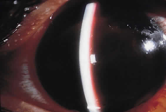
Hyphema is a relatively common complication of ocular trauma (Fig. 1). Bleeding commonly results from tears between the circular and longitudinal muscles of the ciliary body, disrupting the major arterial circle of the iris. These tears produce angle recession. Hyphemas frequently lead to elevated IOP; the larger the hyphema the greater is the likelihood of pressure elevation. The mechanism of glaucoma is obstruction of the trabecular meshwork by trabecular beam swelling, fibrin, and red blood cells. Because of their pliability, red blood cells, in small amounts, pass through normal trabecular meshwork with relative ease.1 However, in larger quantities, especially when the accompanied by fibrin and trabecular swelling, red blood cells may obstruct the meshwork.1,2 Microhyphemas, in which the suspended red blood cells do not form an observable layer, also may lead to elevated IOP. Glaucoma occurs in over 25% of hyphemas filling 50% of the anterior chamber, over 50% of near total hyphemas, and in almost all “black ball” or “eight ball” hyphemas.

Fig. 1. Traumatic hyphema. A. Eighty-five percent hyphema with 50% clot, a smaller clot superiorly, and the margin of pupil visible near the superior edge. B. Near 100% hyphema.

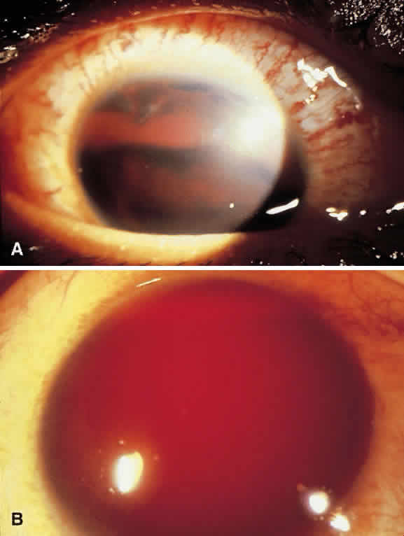
In a “black ball” hyphema, the blood is so concentrated and the anterior chamber so stagnant and hypoxic, that the blood within the anterior chamber appears black (Fig. 2). Such a black ball hyphema may consist of a dumbbell shaped clot filling the anterior chamber, pupil, and posterior chamber. These hyphemas are more difficult to manage and lead to glaucoma through pupillary block as well as trabecular obstruction.4

Fig. 2. Traumatic “black ball” hyphema. Anterior chamber is filled with dense blood and clot.

Optimal treatment for hyphema has not been established. Most clinicians recommend treatment with topical steroids and cycloplegics. Patients are often instructed to keep their heads elevated to promote settling of the red blood cells in the inferior anterior chamber angle, thus improving vision and allowing more of the trabecular meshwork to clear. Bed rest and the avoidance of nonsteroidal anti-inflammatories or other agents with blood thinning properties are recommended to reduce the incidence of rebleeds, which commonly occur in the first 4 days.

Rebleeds often are more severe than the initial bleeding episode and may lead to more serious complications and vision loss. Oral aminocaproic acid and steroids have been shown to reduce the rate of rebleeds. Oral aminocaproic acid, however, may cause nausea, vomiting, and systemic hypotension; thus, its use typically requires inpatient hospitalization. Topical aminocaproic acid may also reduce rebleeding.5 Most hyphemas can be managed on an outpatient basis unless the patient is at high risk of rebleeding and complications.6,7 Such patients include those receiving anticoagulant therapy, children, African Americans, patients who tested positive for sickle cell (including trait), patients with greater than one-third hyphemas, and patients at high risk for noncompliance with therapy or follow-up.

Aqueous suppressants are helpful to control elevated IOP. Miotics are avoided because they may increase inflammation or rebleeding. Latanoprost should also be avoided, given concerns regarding increased inflammation and current lack of evidence that it is effective in this setting. Most patients without pre-existing nerve damage or sickle cell disease or trait may be observed, despite moderately elevated IOPs (up to 35 to 40 mm Hg) for 2 to 4 weeks, with only conservative medical therapy.

Patients with severely or persistently elevated pressures may be treated with paracentesis, anterior chamber washout, clot extraction (either manual or facilitated by a mechanical vitrector), or guarded filtration procedures. Some clinicians routinely perform a guarded filtration procedure at the time of anterior chamber washout to help ensure short-term IOP control. The expectation with this combined procedure is that the filtration surgery will most likely fail in the intermediate future, by which time the trabecular meshwork should have recovered adequate function. Trabeculectomy with iridectomy has been recommended as the initial treatment for total hyphemas, because most require surgery eventually.8 During anterior chamber washout, it is not necessary to remove all of the clot, because it is the circulating red blood cells that obstruct the meshwork, and clot removal may lead to rebleeding and damage to intraocular structures. When necessary, an automated vitrectomy cutting handpiece with irrigating sleeve can be helpful to debulk clots without disturbing the structures to which the clots are attached. Ideally, if the clinical situation allows, surgical procedures should be done on or after the fourth day following the trauma, because the highest incidence of rebleeding is between the second and fourth days. Additionally, by this time, clots may have retracted from adjacent structures, facilitating their removal with reduced surgical manipulation and trauma.

All African-American patients or patients with a family history of sickle cell trait or disease who present with a hyphema or microhyphema must be screened for sickle cell trait. The sickle cell gene is present in approximately 9% of African Americans. In the setting of a hyphema, patients with sickle cell trait alone or sickle cell disease may have clinically significant sickling of red blood cells in the relatively hypoxic and low pH conditions of the anterior chamber. Sickled red blood cells are less pliable and easily obstruct the trabecular meshwork, leading to high IOPs even with remarkably little blood in the anterior chamber.9 In addition, patients with sickle cell trait or disease are at risk of ischemic complications to the optic nerve or retina at relatively low IOPs.10,11 For this reason, it has been suggested that sickle patients with hyphemas or microhyphemas be monitored more closely, and that their IOP be managed aggressively to be maintained below 30 mm Hg at all times, and not be allowed to remain higher than 24 mm Hg for more than 24 hours.

Therapies for elevated IOP secondary to hyphemas in patients with sickle cell disease are similar to those in patients without sickle cell. However, there are some important additional considerations.12 Carbonic anhydrase inhibitors may lead to systemic acidosis, potentially precipitating systemic sickle crises or exacerbating sickling in the anterior chamber with attendant complications. Methazolamide has less potential for this than acetazolamide. The significance of the effects of topical carbonic anhydrase inhibitors on the aqueous in this setting is unknown. Systemic osmotic agents may dehydrate patients and also promote systemic sickling in those so predisposed. Epinephrine may increase anterior chamber acidosis through its ischemic effects; the potential of apraclonidine to do the same is unproven. Brimonidine's effects in this situation have also not been fully described. However, brimonidine is highly selective for α-2 (1800:1 α-2:α-1) thus it has less vasoconstrictive (α-1 mediated) effects compared with the other α agonists. Earlier paracentesis has been recommended in the presence of sickle cell trait or disease.12

Corneal blood staining, in which hemoglobin enters the cornea stroma, is another potential complication of hyphemas. The likelihood of corneal blood staining is correlated with longer duration of blood in the anterior chamber, higher IOP, and reduction in the health of the corneal endothelium.13,14 Corneal blood staining clears eventually, starting peripherally, unless the staining is severe or the cornea has underlying dysfunction. Complete clearing may take 6 months or more. In children under 9 years of age, a dense amblyopia may result. For this reason, persistent hyphemas should be more aggressively managed in pediatric patients.

LENS-RELATED GLAUCOMAS

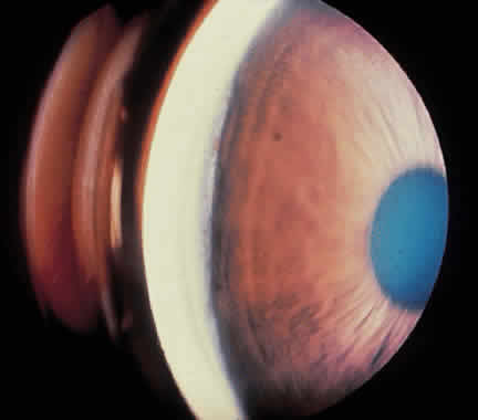
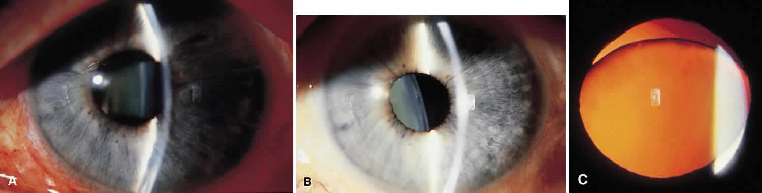
Traumatic dislocation or subluxation of the crystalline lens may lead to pupillary block glaucoma. The loose lens may be free to move anteriorly into the pupil or anterior chamber intermittently, which creates pupillary block (Fig. 3). The loose lens may also produce acute and chronic angle closure by a mass effect, pushing the iris anteriorly. A significantly dislocated lens may allow the vitreous to come forward and cause pupillary block glaucoma. Assessment of lens motility is important in planning glaucoma surgery for any patient with a history of trauma. Occasionally, a lens is noted to be loose at the beginning of surgery, when the supine position leads to the lens's drifting posteriorly or allows the vitreous to come forward around the lens. Remarkably, the lens often re-establishes its original position when the patient is again upright following surgery. Vitreous in the anterior chamber may block the sclerostomy, thus leading to filtration failure.

Fig. 3. Lens-related glaucoma secondary to trauma. A. Shallow anterior chamber secondary to pupillary block caused by loose lens rotating anteriorly at superior pole. B. Following laser peripheral iridotomy, the chamber is deep. Lens has moved posteriorly C. Retroillumination following dilation (after the iridotomy) shows subluxation of lens.

Phacolytic glaucoma may rarely occur following blunt trauma. Significant contusion injury to the lens may lead to massive swelling of the lens and leakage of lens proteins. Aqueous outflow is obstructed by these proteins and by macrophages that engulf them.3 Swollen lenses may also lead to phacomorphic glaucoma with angle closure. Lens particle glaucoma may occur when the lens capsule is torn by sharp, or occasionally by blunt, trauma lead to trabecular meshwork obstruction by lens fragments.

CHEMICAL INJURIES

Exposure to strongly basic solutions may denature the proteins in the sclera and lead to scleral shrinkage with an acute rise in IOP. Such pressure may drop transiently, only to elevate severely once again following release of prostaglandins.15,16 In rabbits, this secondary pressure rise was blocked by therapy with oral aspirin or indomethacin.16 These glaucomas are treated with aqueous suppressants and topical steroids. However, associated severe corneal injuries, which often present the risk of full thickness corneal melts, may limit the use of steroids.

CAROTID CAVERNOUS AND DURAL FISTULAS

Trauma may tear the internal carotid artery within the cavernous sinus, elevating the local venous system pressure to arterial pressure level.17,18 Tears in the branches of the internal and external carotid arteries may lead to communications with the dural sinus. A sign of this is the presence of dilated vessels beneath the conjunctiva, especially when these do not blanch with epinephrine exposure. Blood may be seen in Schlemm's canal on gonioscopy. Superior vena cava syndrome and thrombosis of an orbital vein or of the cavernous sinus can produce similar findings.

The aqueous outflow pathway may be summarized as follows: anterior chamber → Schlemm's canal → aqueous veins → episcleral veins → anterior ciliary veins → superior ophthalmic vein → cavernous sinus → internal jugular vein. Some conjunctival veins also carry aqueous through the facial vein to the external jugular vein.

Goldmann's equation, which describes the relation of IOP to aqueous flow and episcleral venous pressure, is Po = F/C + Pev; where Po is IOP, F is aqueous production, C outflow facility, and Pev is episcleral venous pressure. This theoretical model has shown good correlation with clinical experience, animal research, and human cadaver experiments. The equation predicts that increases in episcleral venous pressure lead directly to increases in IOP. Normal episcleral venous pressure is between 9 and 10 mm Hg.

Arterialization of the outflow pathways of the eye also may lead to ocular ischemia and neovascular glaucoma.19 Increased venous pressure may lead to choroidal effusions and suprachoroidal hemorrhages with shallowing of the anterior chamber and secondary angle closure glaucoma. Because the mechanism of the angle closure glaucoma is not pupillary block, iridectomy is not helpful.

Treatment of the underlying cause of increased venous pressure is ideal but not always possible. Embolization of carotid-cavernous fistulas carries significant risk of central nervous system vascular accident; many dural fistulas close spontaneously. Aqueous suppressants often are helpful in controlling IOP. Miotics and argon laser trabeculoplasty are not useful given the typically normal facility of outflow. Guarded filtration surgery (trabeculectomy) is effective in that it bypasses the elevated episcleral venous pressure, but it has a higher incidence in these patients of serous and hemorrhagic choroidal detachments. Tight closure of the scleral flap covering the entrance to the anterior chamber is recommended to reduce the incidence of serous and hemorrhagic choroidal effusions. Postoperative suture release can then be used to titrate the reduction in IOP, minimizing sudden pressure changes and the potential for choroidal hemorrhage and effusions. Some clinicians advocate prophylactic “scleral windows” over the ciliary body, either as flaps or by thinning the sclera to about one half its normal thickness, to facilitate drainage of choroidals and prevent large or expulsive choroidal detachments. These are typically placed in the inferior quadrants.

Back to Top
LATE POSTTRAUMATIC GLAUCOMA

GHOST CELL

Ghost cells are degenerate red blood cells whose leaky membranes have allowed much of the cell's hemoglobin to escape. Residual, degenerated hemoglobin precipitates on inner cell walls in the form of Heinz bodies. These spherical ghost cells are much less pliable than red blood cells and cannot pass through the trabecular meshwork. Ghost cells in the anterior chamber result in elevated IOP much more readily than red blood cells.20

Formation of ghost cells requires sequestration of red blood cells in the vitreous. After several weeks, degenerated cells migrate into the anterior chamber through a defect in the vitreous face, surgical or traumatic, and obstruct the trabecular meshwork. Tan or khaki cells are seen in the anterior chamber, anterior vitreous, the trabecular meshwork, and may also form a pseudohypopyon. Paracentesis may confirm the diagnosis. Phase contrast microscopy, cell block, or millipore filter preparations reveal ghost cells.21,22 Specimens may be submitted to laboratories using the same methods as for cerebrospinal fluid.

Hemolytic glaucoma occurs when the contents of ruptured red blood cells are ingested by macrophages, which then obstruct the meshwork. However, unlike ghost cell glaucoma, hemolytic glaucoma is typically seen in the first days to weeks after initial hemorrhage.

Aqueous suppressants are the most useful agents in attempting to control IOP. Miotics are rarely helpful because the meshwork is obstructed. Argon laser trabeculoplasty is contraindicated. Anterior chamber lavage with balanced salt solution may resolve the glaucoma. If unsuccessful, or too great a reservoir of ghost cells remains in the vitreous, anterior chamber washout combined with total vitrectomy, removing as much hemorrhagic material as possible, is usually effective in resolving the glaucoma.23

SIDEROSIS: BLOOD AND FERROUS FOREIGN BODIES

Hemosiderotic glaucoma, in which iron-containing blood breakdown products stain and poison the cells of the trabecular meshwork, occurs much later, often years after the hemorrhage. Iron-containing foreign bodies may also lead to loss of trabecular meshwork function. Iron may stain Descemet's membrane and lead to a rusty discoloration of the anterior capsule of the lens. Additionally, iris heterochromia may be a clue to the diagnosis. Gonioscopy and thorough, dilated fundus examination often reveals the foreign body. Metallic foreign bodies may be detected by radiographs, computed tomography scan, and ultrasound. Occasionally, ultrasound biomicroscope (UBM) may allow detection and localization of foreign bodies in the anterior chamber angle and ciliary body. Removal of the iron-containing foreign body is often indicated without taking the glaucoma into account, because attendant siderosis also may be toxic to the retina.

ANGLE RECESSION

Angle recession glaucoma is an important late complication of ocular trauma (Figs. 4 and 5). Angle recession is seen in most patients in whom trauma has caused a hyphema.24,25 IOP may remain normal for years to decades and then become severely elevated. Patients must be strongly advised of the need for lifelong, regular follow-up following significant ocular trauma. Elevated IOP is more likely to occur in eyes with 180 degrees or more of angle recession.24,26 Angle recession glaucoma may occur in 6% to 20% over a 10-year period.25 Angle recession must be considered in chronic unilateral glaucomas. Patients often do not recall any long past trauma. Clues to the existence of such earlier trauma include facial and corneal scars, pupillary sphincter tears, iris transillumination, iridodonesis, phacodonesis, Vossius' ring, dark brown or black deposits in the inferior angle recess, and localized anterior synechiae as a sign of earlier intraocular inflammation, especially at the 6-o'clock position. With time, angle recession may scar, becoming less evident by gonioscopy in apparent severity and extent. Affected areas continue to have nonfunctioning meshwork, secondary to scarring or their closure by a Desçemet-like membrane.27–29 Interestingly, the fellow eye is at significantly increased risk of development of open angle glaucoma.

Fig. 4. Traumatic angle recession. Temporally, for two clock hours, a widened band of the ciliary body has been exposed. This is in contrast to the remainder of the angle, where only the anterior portion of the ciliary body is visible.

Fig. 5. Traumatic angle recession. A. Normal anterior chamber angle. A line parallel to the visual axis through the trabecular meshwork crosses the tip of the first ciliary process. B. Traumatic angle recession. Iris insertion is moved posteriorly with rotation of the ciliary body. A line parallel to the visual axis through the trabecular meshwork passes internal to the retracted ciliary body processes. (Courtesy of Ralph C. Eagle, Jr.)

Angle recession glaucoma may respond to aqueous suppressants. Miotics and argon laser trabeculoplasty are rarely effective. Long-term success rates for trabeculectomy are lower than for primary open angle glaucoma; some surgeons use antimetabolites on primary procedures for this reason.

CHRONIC ANGLE CLOSURE GLAUCOMAS

Chronic angle closure glaucoma may develop following trauma through various mechanisms. Perforating trauma may lead to shallowing of the anterior chamber, which may lead to anterior synechiae in the setting of inflammation. Bleeding and inflammation associated with blunt trauma may create anterior and posterior synechiae. Lens subluxation or swelling may also lead to acute and chronic angle closure. Some clinicians advocate surgical or laser intervention to clear synechiae within the first 6 months of their formation, but many clinicians are not convinced that this alters the long-term clinical course.

SCHWARTZ'S SYNDROME

Schwartz's syndrome is elevated IOP secondary to obstruction of the trabecular meshwork by photoreceptor outer segments.30 These photoreceptor outer segments are released from a chronic retinal detachment through a tear in the retina. Retinal detachments are often low lying and peripheral and so may be easily overlooked. The photoreceptor outer segments may be mistaken for white blood cells and the glaucoma erroneously diagnosed as uveitic. Pressures may fluctuate widely depending on the balance of trabecular meshwork outflow obstruction and increased outflow through retinal detachment. The retinal pigment epithelium has a pumping function that normally evacuates subretinal fluid, keeping the retina firmly attached to the pigment epithelium. In the case of a retinal detachment, this pumping function helps to lower IOP. This can very rarely lead to iris retraction syndrome, in which patients with Schwartz's syndrome and extensive posterior synechiae show iris retraction when placed on therapy that includes aqueous suppressants. The theory is that the reduced inflow from aqueous suppression, coupled with the reduced flow into the anterior chamber and increased outflow through the retinal detachment, leads to hypotony and an exceptionally deep anterior chamber.

CYCLODIALYSIS CLEFT CLOSURE

Traumatic cyclodialysis clefts divert aqueous away from the trabecular meshwork and may lead to hypotony. The unused trabecular meshwork becomes less permeable to aqueous. Months or years later, spontaneous closure of the cyclodialysis cleft may lead to dramatic elevations of IOP. Patients present with sudden onset of ocular pain, headaches, and reduced vision. Slit lamp and gonioscopic evaluation differentiate this entity from the similar presentation of acute angle closure glaucoma. Aqueous suppressants are often effective in reducing IOP on a long-term basis, or until the trabecular meshwork recovers and better outflow facility resumes. Miotics are avoided because they may reopen cyclodialysis clefts. An exception to this is a long-standing cleft with good IOP control that was well tolerated. In this situation, immediate use of a strong miotic, such as pilocarpine 4%, may open and re-establish the cleft. Occasionally, filtering surgery may be necessary in patients with persistently elevated pressures.

EPITHELIAL DOWNGROWTH AND STROMAL INGROWTH

Perforating injuries create a pathway for epithelial and fibrous connective tissue to grow into the eye.31 Membranes, often with scalloped edges, may proliferate across anterior chamber structures including the iris and corneal endothelium (Fig. 6). These membranes cover the trabecular meshwork and lead to reduced outflow. The membranes may be identified by argon laser treatment over affected portions of the iris. The laser creates white, rather than brown, spots.

Fig. 6. Epithelial ingrowth. An epithelial membrane covers the anterior iris and the anterior chamber angle recess. (Courtesy of Ralph C. Eagle, Jr.)

Treating these conditions is extremely difficult. Filtering surgeries often fail quickly, even with antimetabolite use, because internally proliferating membranes close the sclerostomy. Tube shunts have been advocated, but these may also fail. Cryotherapy to the cornea and iris has been advocated to destroy the advancing membranes, but may lead to damage to other ocular structures. In selected cases, meticulous surgical removal of the involved areas of iris with membrane peeling from the cornea may be successful.

Back to Top
REFERENCES

1. Horven I: Erythrocyte resorption from the anterior chamber of the human eye. Acta Ophthalmol 41:402, 1963

2. Horven I, Myhre E: Erythrocyte resorption from the anterior chamber of the eye in rabbits. Acta Ophthalmol 41:385, 1963

3. Epstein DL, Hashimoto JM, Grant WM: Serum obstruction of aqueous outflow in enucleated eyes. Am J Ophthalmol 86:101, 1978

4. Parrish R, Bernardino V: Iridectomy in the surgical management of eight-ball hyphema. Arch Ophthalmol 100:435, 1982

5. Crouch ER Jr, Williams PB, Gray MK et al: Topical aminocaproic acid in the treatment of traumatic hyphema. Arch Ophthalmol 115:1106, 1997

6. Kennedy RH, Brubaker RF: Traumatic hyphema in a defined population. Am J Ophthalmol 106:123, 1988

7. Shiuey Y, Lucarelli MJ: Traumatic hyphema: Outcomes of outpatient management. Ophthalmology 105:851, 1998

8. Weiss JS, Parrish RK, Anderson DR: Surgical therapy of traumatic hyphema. Ophthalmic Surg 14:4:343, 1983

9. Goldberg MF, Tso MOM: Sickled erythrocytes, hyphema, and secondary glaucoma: VII. The passage of sickled erythrocytes out of the anterior chamber of the human and monkey eye: Light and electron microscopic studies. Ophthalmic Surg 10:89, 1979

10. Michelson PE, Pfaffenback D: Retinal arterial occlusion following ocular trauma in youths with sickle-trait hemoglobinopathy. Am J Ophthalmol 74:3:494, 1972

11. Sorr EM, Goldberg RE: Traumatic central retinal artery occlusion with sickle cell trait. Am J Ophthalmol 80:4:648, 1975

12. Goldberg MF: The diagnosis and treatment of secondary glaucoma after hyphema in sickle cell patients. Am J Ophthalmol 87:43, 1979

13. Brodrick JD: Corneal blood staining after hyphema. Br J Ophthalmol 56:589, 1972

14. Beyer TL, Hirst LW: Corneal blood staining at low pressures. Arch Ophthalmol 103:654, 1985

15. Paterson CA, Pfister RR: Intraocular pressure changes after alkali burns. Arch Ophthalmol 91:211, 1974

16. Paterson CA, Pfister RR: Prostaglandin-like activity in the aqueous humor following alkali burns. Invest Ophthalmol 14:177, 1975

17. Hernderson JW, Schneider RC: The ocular findings in carotid-cavernous fistula in a series of 17 cases. Am J Ophthalmol 48:51:585, 1959

18. Phelphs CD, Thompson HS, Ossoinig KC: The diagnosis and prognosis of atypical carotid-cavernous fistula (red-eye shunt syndrome). Am J Ophthalmol 93:423, 1982

19. Weiss DI, Shaffer RN, Nehrenberg TR: Neovascular glaucoma complicating carotid-cavernous fistula. Arch Ophthalmol 69:304, 1963

20. Campbell DG, Simmons RJ, Grant WM: Ghost cells as a cause of glaucoma. Am J Ophthalmol 81:441, 1976

21. Cameron JD, Havener VR: Histologic confirmation of ghost cell glaucoma by routine light microscopy. Am J Ophthalmol 96:251, 1983

22. Summers CG, Lindstrom RL, Cameron JD: Phase contrast microscopy. Diagnosis of ghost cell glaucoma following cataract extraction. Surv Ophthalmol 28:342, 1984

23. Singh H, Grand MG: Treatment of blood-induced glaucoma by trans pars plana vitrectomy. Retina 1:255, 1981

24. Blanton FM: Anterior chamber angle recession and secondary glaucoma. A study of the aftereffects of traumatic hyphemas. Arch Ophthalmol 72:39, 1964

25. Kaufman JH, Tolpin DW: Glaucoma after traumatic angle recession: a ten-year prospective study. Am J Ophthalmol 78:4:648, 1974

26. Tonjum AM: Intraocular pressure and facility of outflow later after ocular contusion. Acta Ophthalmol 46:886, 1968

27. Reese AB: Deep-chamber glaucoma due to the formation of a cuticular product in the filtration angle. Am J Ophthalmol 27:11:1193, 1944

28. Lauring L: Anterior chamber glass membranes. Am J Ophthalmol 68:308, 1969

29. Waring GO, Laibson PR, Rodrigues M: Clinical and pathologic alteration of Descemet's membrane: With emphasis on endothelial metaplasia. Surv Ophthalmol 18:5:325, 1974

30. Matsuo N, Takabatake M, Ueno H et al: Photoreceptor outer segment in the aqueous humor in rhegmatogenous retinal detachment. Am J Ophthalmol 101:673, 1986

31. Allen JC: Epithelial and stromal ingrowths. Am J Ophthalmol 65:2:179, 1968

Back to Top