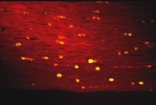
Fig. 3.
Acanthamoeba
in cornea stained with calcifluor white and imaged with ultraviolet fluorescence microscopy. (Photomicrograph courtesy of Dr. Morton Smith.)