|
|
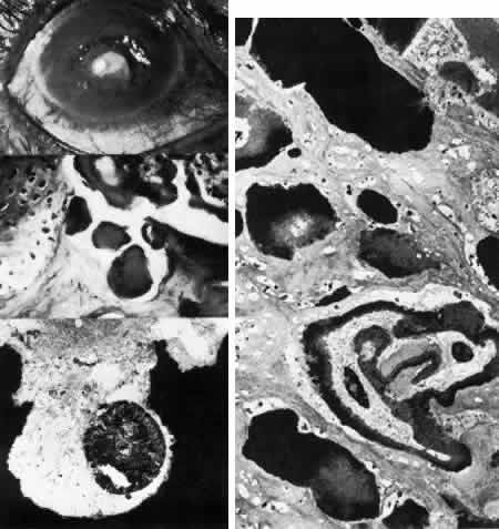
| Fig. 27, Spheroid (keratinoid) degeneration. Top left. Clinically, numerous spheroidal deposits appear as opacities over the anterior stroma. Middle left. Histologic section reveals numerous densely staining spherules beneath the distorted epithelium and within the superficial stroma (hematoxylin-eosin. × 250). Right. Survey transmission electron micrograph shows spheroidal deposits as extracellular accumulations of electron-dense material with variably crystalline structure. Lipid substances and blood vessels are also evident (× 5000). Bottom left. High-magnification transmission electron micrograph of a spheroidal deposit shows variable electron density with a crystalline fragment similar to calcium (× 40,000). |