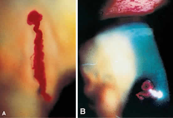
Fig. 2.
A.
High-magnification photograph of a filament showing a mucous filament based on a patch of abnormal epithelium.
B.
Slit-lamp photograph of mucous plaques on the corneal surface in a patient with herpes zoster ophthalmicus.