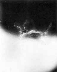
Fig. 11
Macrophotograph of portion of a dendritic ulcer by retroillumination. Note opaque heaped-up cells along the ulcer margin. (Magnification × 10) (Courtesy of Mr. N. Brown)